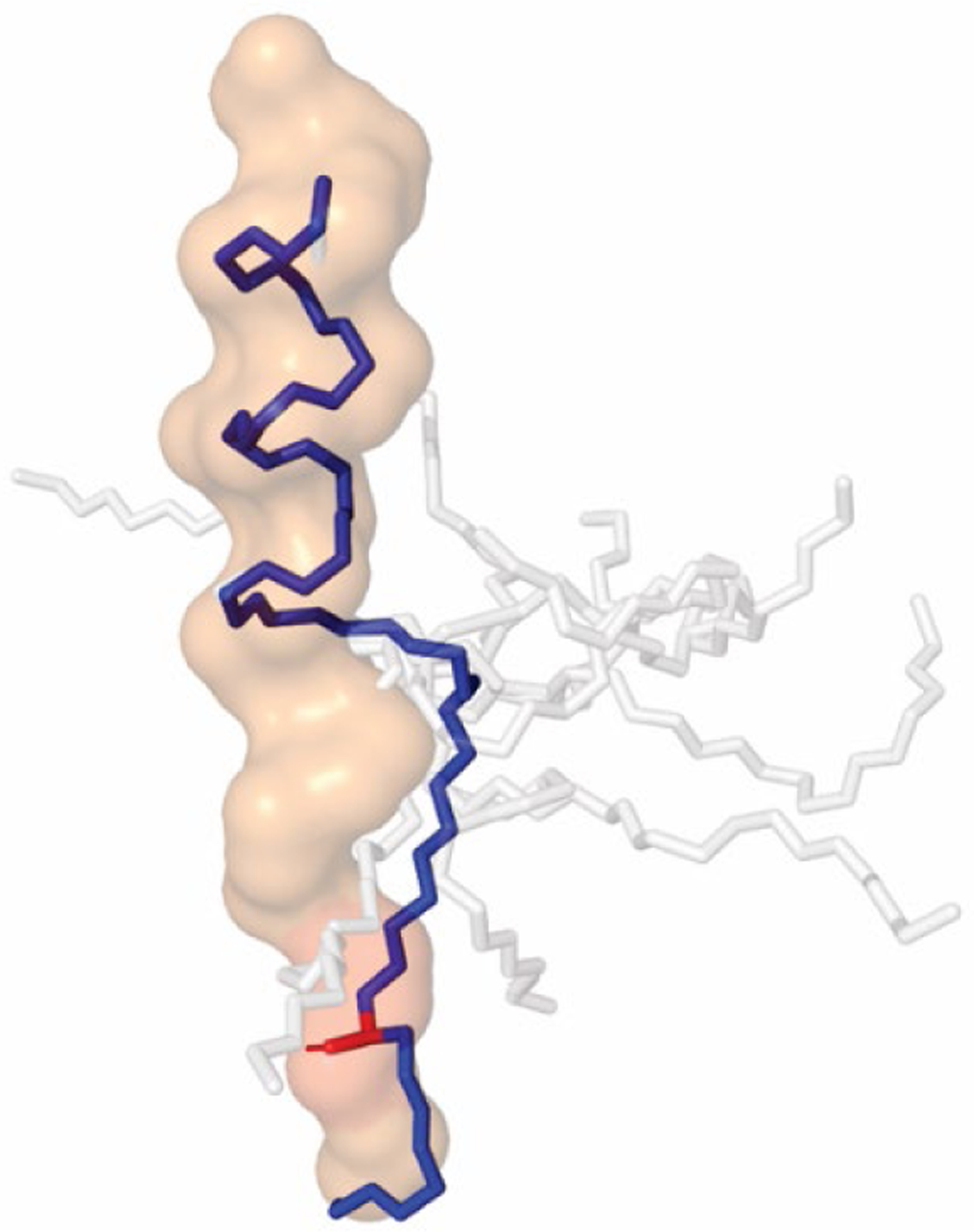
Figure 4: Solution conformation of designed HPI-TSA conjugate 6 resembles that of APP TMD substrate bound to γ-secretase.

NMR constraints were used to determine low-energy conformations of 6. The top 10 conformers are shown as sticks, with the conformer closest to that of the bound substrate in blue. Structure rendered in Pymol using PDB file 6IYC for bound APP substrate, shown as surface outline. Transition-state mimicking hydroxyl group (red) of 6 overlaps with the scissile amide bond in the extended region of APP substrate when the helical moiety is aligned with the helical region of bound APP substrate.
