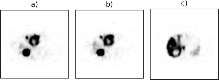
Fig. 2.
SPECT images of a 69-year-old male with neuroendocrine tumor in the pancreas. The patient was diagnosed with metastatic spread to the liver, which was treated with a 166Ho radioembolization procedure in the whole liver (prescribed activity 9900 MBq). a166Ho-DI and b166Ho-only acquisition images have been, independently and blindly, presented to the nuclear medicine physicians for the qualitative assessment. c99mTc image acquired during the DI protocol where an additional 50 MBq of 99mTc-colloid was administered

