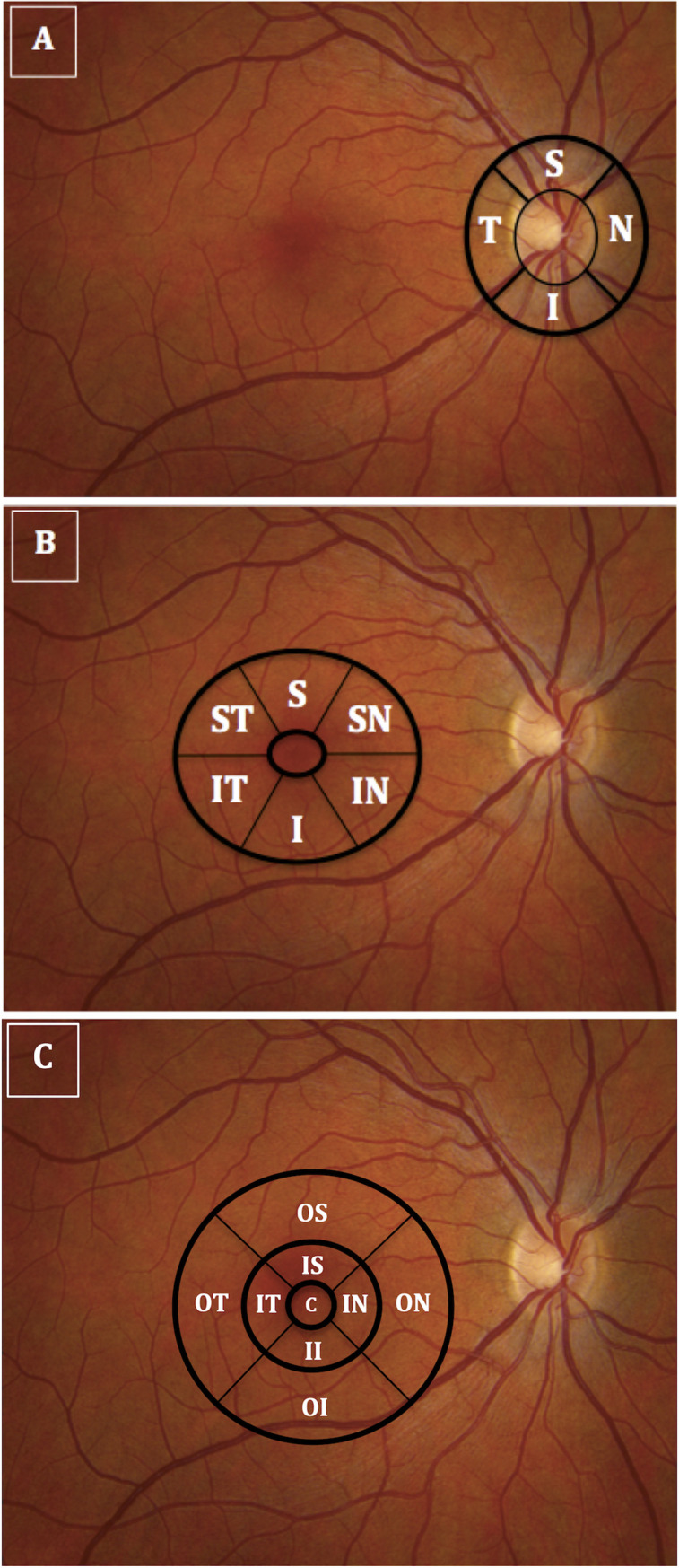
Fig 1. Illustrates optical coherence tomography imaging of the peripapillary and macular areas.

(A) The thicknesses of the 4 retinal nerve fiber layer quadrants (temporal, superior, nasal, inferior); (B) ganglion cell-inner plexiform layer sectors (superior-temporal, superior, superior-nasal, inferior-nasal, inferior, inferior-temporal; (C) macular full-thickness sectors (center, inner-superior, outer-superior, inner-inferior, outer-inferior, inner-temporal, outer-temporal, inner-nasal, outer-nasal) were measured using peripapillary and macular circular scans centered on the disc and on the fovea, respectively).
