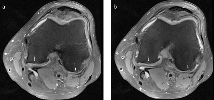
Figure 4. a, b.
Consecutive axial fat-suppressed proton density-weighted images (a) and (b) from MRI study of the right knee of a 49-year-old woman with a PHMM RL tear demonstrates ICRS Grade 4 chondral lesions of the medial and lateral FPFC. The white arrows show subchondral cyst like and edema-like lesion in the medial and lateral FPFC. There are ICRS Grade 4 cartilage lesions noted. The cartilage is severely abnormal extending down through the subchondral bone.

