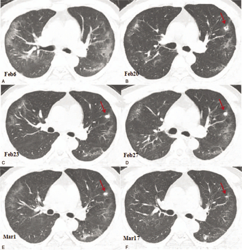
Figure 1.

Computed tomography (CT) images showing the changes of the cavity in the anterior segment of the left upper lobe. (A) A CT image obtained on February 6, 2020 showing the ground glass opacities and linear opacities in the lungs predominantly distributed in the peripheral third of the lungs. (B) A CT image obtained on February 20, 2020 showing the reduced ground glass opacities area. A small cavity was found in the anterior segment of the left upper lobe with a size of 9.3 × 6.7 mm. (C) A CT image obtained on February 23, 2020 showing consolidation of the small cavity in the anterior segment of the left lobe with a size of 7.8 × 6.3 mm (arrow); (D) A CT image obtained on February 27 showing consolidation of the cavity in the upper segment of the left upper lobe with the size slightly reduced to 6.5 × 5.5 mm (arrow); (E) A CT image obtained on March 1, 2020 showing the reduced cavity in the anterior segment of the left upper lobe, with a size of 5.0 × 4.5 mm (arrow). (F) A CT image obtained on March 17, 2020 showing that the ground glass opacities in the lungs were almost completely absorbed. The solid nodule in the anterior segment of the left upper lobe had significantly reduced in size, with a size of 2.5 × 1.5 mm (arrow).
