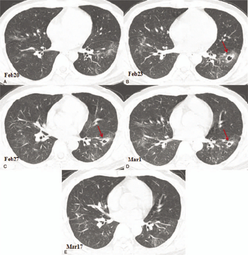
Figure 2.

Chest computed tomography (CT) images showing cavitary changes in the inner anterior basal segment of the left lower lobe. (A) A CT image obtained on February 20, 2020 showing the ground glass opacities in the lungs. (B) Compared with the CT image obtained on February 20, 2020, the chest CT image of February 23, 2020 showing a new cavity with a partition in the anterior basal segment of the left lower lobe with a size of 15.0 × 13.0 mm (arrow). (C) A chest CT image showing that the anterior basal cavity in the left lower lobe is slightly reduced, with a size of 12.0 × 11.0 mm (arrow). (D) A chest CT image obtanined on March 1st showing that the anterior basal cavity in the lower left lobe is further reduced, the size is 11.0 × 10.0 mm, and liquid-gas level appears (arrow). (E) A chest CT image obtained on March 17th showing the cavity in the inner anterior basal segment of the left lower lobe was disappeared.
