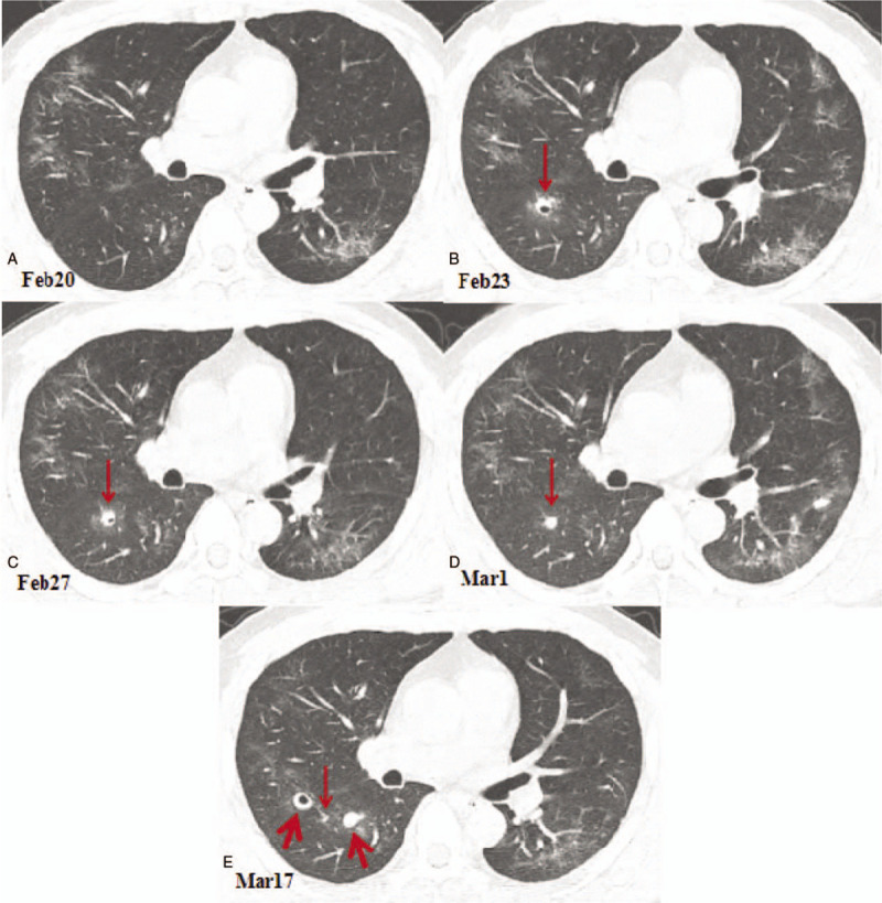
Figure 3.

Chest computed tomography (CT) images showing cavitary change in the dorsal segment of the right lower lobe. (A) A CT image obtained on February 20, 2020 showing the ground glass opacities in the lungs. (B) Compared with the CT image obtained on February 20, 2020, the chest CT image obtained on February 23, 2020 showing a new cavity in the dorsal segment of the right lower lobe with a size of 8.2 × 7.1 mm (arrow). (C). A chest CT image obtained on February 27th showing a part of cavity in the dorsal segment of the right lower lobe was condensed, with a size of 7.7 × 6.3 mm (arrow). (D).A chest CT image obtained on March 1st showing that the cavity of the lower right lobe was completely consolidated and shrunk to a size of 6.1 × 5.0 mm (arrow). (E) A chest CT image obtained on March 17th showing that the lesion in the dorsal segment of the right lower lobe was almost completely absorbed, leaving only a punctate shadow (thin arrow). On the left side of the lesion, a new solid nodule with a size of 9.8 × 8.5 mm appears at the dorsal lobe of the right lower lobe (thick arrow); on the right side of the lesion, a new cavity appears in the posterior segment of the right upper lobe with a size of 11.5 × 10.3 mm (thick arrow).
