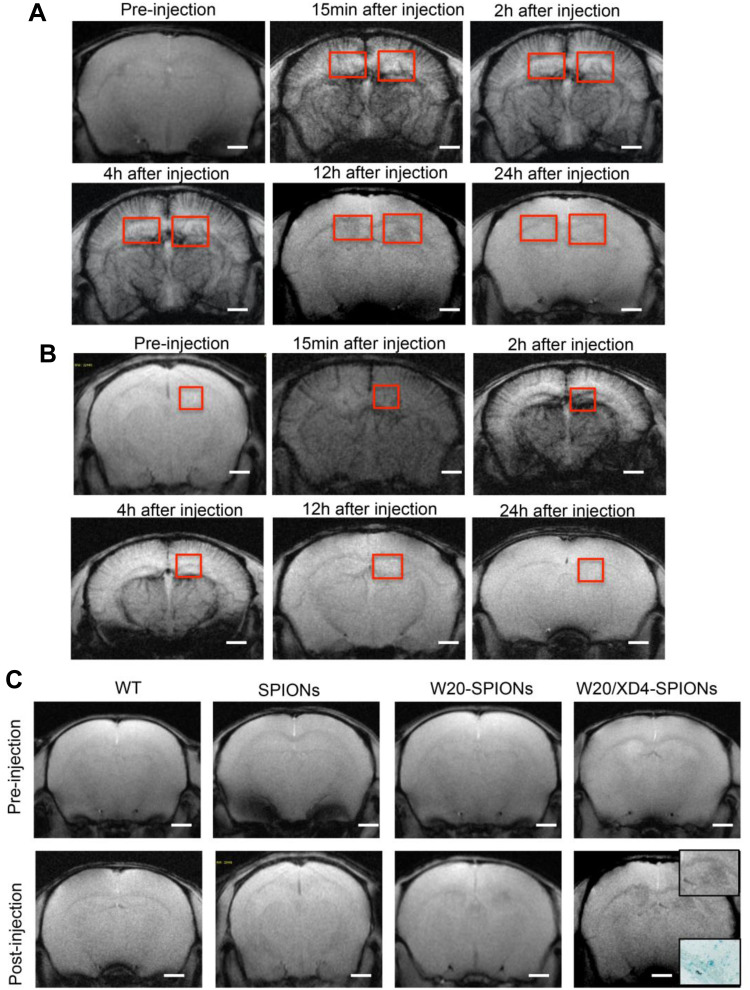
Figure 6.
W20/XD4-SPIONs exhibited remarkable MR signal in AD mouse brains. (A and B) In vivo T2*-weighted images of W20/XD4-SPIONs distribution in AD (A) and WT (B) mouse brains were captured pre- and 15 min, 2 h, 4 h, 12 h and 24 h post intravenous injection of W20/XD4-SPIONs. (C) In vivo T2*-weighted images of the probe distribution in AD mouse brains after intravenous injection of W20/XD4-SPIONs, W20-SPIONs and SPIONs. Boxed regions are shown at higher magnification or stained by the Prussian blue. Scale bar, 1 mm.

