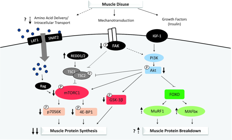
FIGURE 1.
Molecular mechanisms of skeletal muscle disuse atrophy. Declines in muscle protein synthesis (postabsorptive and postprandial) and a possible early increase in muscle protein breakdown underlie the loss of muscle mass with disuse. Intracellular mechanisms responsible for these changes in protein turnover may include impairments in delivery and intracellular transport of amino acids, declines in mechanotransduction, and insulin resistance specific to protein metabolism leading to downstream attenuation anabolic signaling (mTOR-dependent and -independent pathways) and a possible increase in ubiquitin-mediated proteolysis. Solid arrows indicate consistent findings, while broken arrows suggest potential mechanisms. FAK, focal adhesion kinase; FOXO, forkhead box O; GSK-3β, glycogen synthase kinase 3β; IGF-1, insulin-like growth factor 1; LAT1, L-type amino acid transporter 1; MAFbx, muscle atrophy F-box; mTORC1, mammalian target of rapamycin complex 1; MuRF1, muscle ring finger 1; p70S6K, p70 ribosomal protein S6 kinase; PI3K, phosphoinositide-3-kinase; REDD1/2, regulated in development and DNA damage 1/2; SNAT2, sodium-coupled neutral amino acid transporter 2; TSC1, tuberous sclerosis complex 1; TSC2, tuberous sclerosis complex 2; 4E-BP1, 4E binding protein 1.

