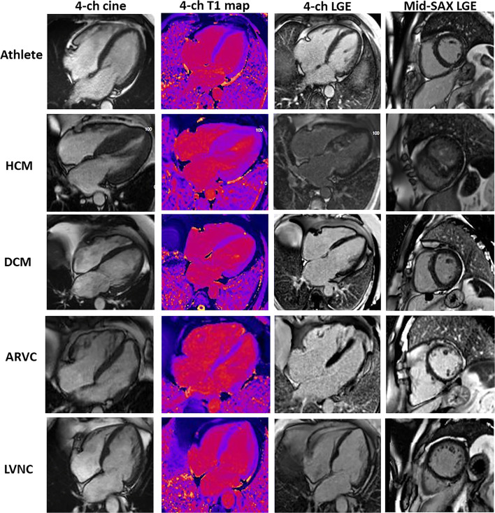
Fig. 2.
CMR findings in the athlete’s heart and mimics. Images show (left to right) 1) an end-diastolic 4-chamber cine; 2) a 4-chamber T1 map; 3) a 4-chamber LGE; 4) a mid-ventricular short axis LGE; in the following conditions (top to bottom): a athlete: demonstrating enlarged cardiac chambers with normal wall thickness. Native T1 map and 4-chamber LGE view are normal. There is inferior RV insertion point LGE, a normal finding when it is just a gram or so, as here. b HCM: predominantly septal LVH. Patchy elevated native T1 with patchy mid-wall LGE seen in hypertrophied myocardium and (here) the papillary muscles. c DCM: LV dilatation with normal native T1 map and no LGE. d ARVC: biventricular ARVC. Elevated native T1 is appreciated within the RV wall as well as extensive biventricular LGE. e LVNC: non-compaction of the LV. Native T1 map is normal and there is subtle LGE seen in the 4-chamber image within the apical cap. This appearance has to be interpreted with context, particularly of ethnicity and athleticism

