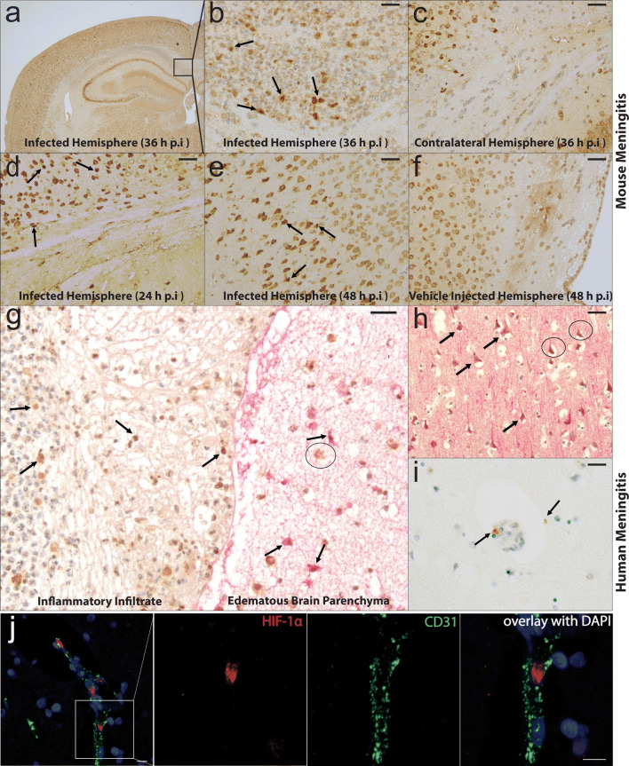
Fig. 1.
Induction of HIF-1α in mouse and human brain tissue samples from pneumococcal meningitis. a Immunohistochemistry of brain tissue 36 h post-S. pneumoniae infection in intracerebrally infected mouse model shows positive HIF-1α in several inflammatory and neural cells. b Zoomed area from a in the cortex region of the infected mouse brain shows significant nuclear HIF-1α (black arrows) indicating its activation along with positive staining in the granulocytic infiltrate. c Non-infected hemisphere of the same mouse at 36 h post infection shows no specific staining for HIF-1α signal. d Positive staining in the cortex region (black arrows) was also observed at 24 h post-intracerebral infection, which was also observed at e 48 h post-infection in the intracisternally infected mice. f Control mice that were intracerebrally injected with 0.9% NaCl did not show any positive staining in the same region. g Brain sections from human pneumococcal meningitis patients double stained for HIF-1α (brown) and GFAP (red) show nuclear staining for HIF-1α (black arrows) in lymphocytes of the inflammatory infiltrate (g, left) and in the edematous brain tissue comprising GFAP positive reactive astrocytes (g, right), where macrophages showed cytoplasmic staining (black circles). h Neurons labeled with MAP2 (red) were also co-stained with HIF-1α (brown) in the cortex but mostly the pyknotic ones and not the vital neurons (black circles). i Positive HIF-1α staining could also be observed in vessel associated cells in the cortex region. j Activated HIF-1α indicated by nuclear staining (red) could be observed in brains ECs from pneumococcal meningitis patients by immunofluorescence staining using CD31 as an endothelial marker. Scale bar b–f is 20 µm, g–i is 25 µm, j left most image 20 µm and the zoomed images 10 µm. Mouse brain stainings are representative of three mice each at 24, 36, 48 h post-infection and two control mice at 48 h post- vehicle injection. Human pneumococcal meningitis stainings are representative of 4 cases outlined in Table 1

