Fig. 4.
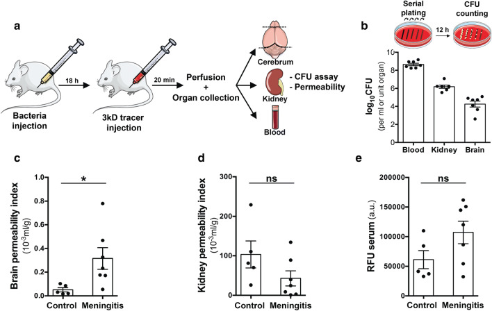
Extravasation of S. pneumoniae and neurovascular permeability in vivo in a hematogenous meningitis model. a Setup showing the murine infection model with intraperitoneal injection of 0.5 × 10⁵ bacteria (in 100 μl PBS, D39 strain). 18 h post-infection mice were injected with a 3kD TMR tracer (i.p), circulated for 20 min followed by anesthesia, transcardial PBS perfusion and collection of blood, brain, and kidney. b Homogenized organs or blood were plated and cultured overnight to obtain the bacterial counts using the CFU assay. High CFU values in blood indicated the hematogenous presence of bacteria with extravasation of bacteria evident in the brain (0.01%). The bacterial counts in the kidney were 100-fold higher than brain reflecting higher permeability of the kidney vasculature. Healthy control mice did not have any bacterial growth on the plates. c Homogenized hemi-brain, kidney and blood were utilized to obtain dextran permeability by measuring fluorescence intensity on a microplate reader. A significant elevation in vascular permeability of infected mice was observed compared to healthy controls in the brain indicating blood–brain barrier breakdown. d Vascular permeability of kidney was not altered upon infection. e Serum fluorescence values (arbitrary units—a.u.) indicate equivalent tracer absorption between healthy and infected mice. (N = 5–7/group, *p < 0.05, 2-tailed unpaired t test)

