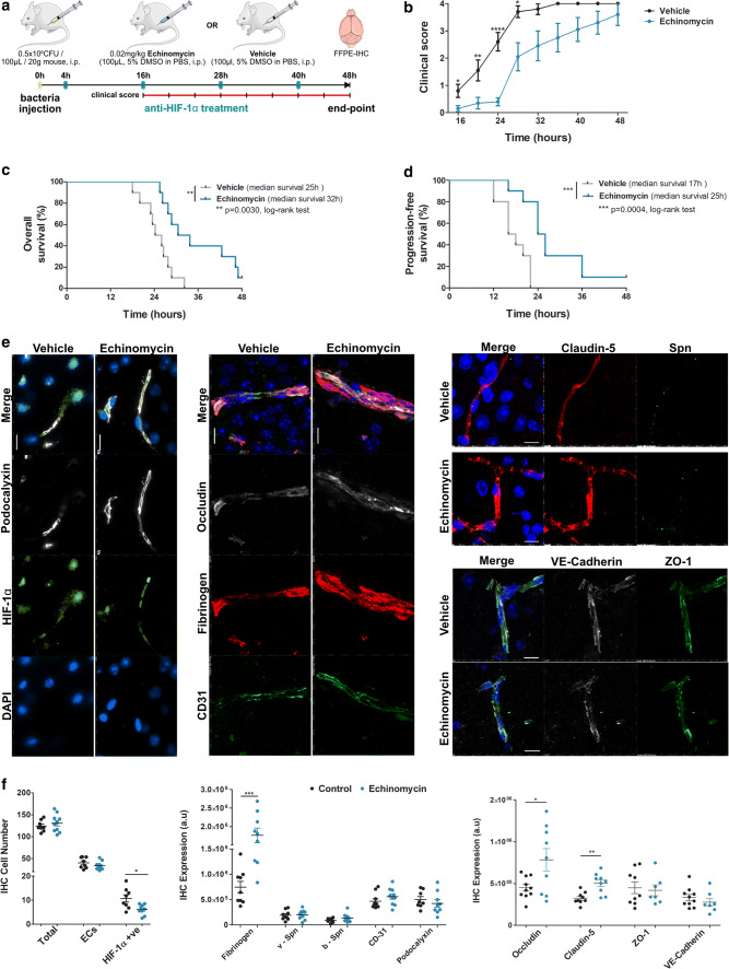
Fig. 8.
Improved survival and blood–brain barrier function post pneumococcal infection in mice by anti-HIF-1α treatment using echinomycin. a Schematic depicting treatment protocol of mice infected with S. pneumoniae (D39). Mice were infected i.p., followed by anti-HIF-1α treatment with echinomycin or just vehicle every 12 h starting 4 h post-infection (blue tick marks). Clinical scores were taken every 4 h starting 16 h post-infection (red line with tick marks) and study terminated at 48 h post-infection and brain tissues collected upon animal death or at the end point when the animals were sacrificed. b Clinical scores indicate improved symptoms in echinomycin-treated mice compared to vehicle-treated animals starting 16 h post-infection that were progression free (below score 1) up to 24 h (mean ± SEM at each time, N = 10/group, ****p < 0.0001, **p < 0.01, *p < 0.05 by 2-tailed unpaired non-parametric Mann–Whitney t test at each time point. c Echinomycin-treated mice showed significantly improved overall survival percentage by Kaplan–Meier analysis with the median survival at 32 h compared to 25 h in vehicle-treated mice (**p < 0.01, log-rank test, N = 10/group). d Kaplan–Meier analysis also indicated a significant improvement in progression-free survival in echinomycin-treated animals (median survival 25 h) with no clinical symptoms up to 24 h (b) compared to the vehicle group (median survival 17 h; ***p < 0.001, log-rank test, N = 10/group). e Immunofluorescence staining for HIF-1α, BBB permeability and junctional markers in the echinomycin and vehicle groups at the survival end point. Left panel shows that echinomycin treatment leads to a reduction in HIF-1α-positive nuclei including in ECs co-stained for podocalyxin, a vascular marker which was unchanged by the treatment. Middle panel displays reduced vascular permeability to fibrinogen in the echinomycin group as indicated by stronger intravascular signal compared to the vehicle-treated mice. Increased expression of tight junction proteins—occludin (middle panel) and claudin-5 (right top panel)—in echinomycin-treated mice indicate improved BBB function. Tight junction-associated ZO-1, adherens junction marker VE-Cadherin, and endothelial cell adhesion molecule CD31 were unchanged (middle, bottom right panel). There was also no difference in S. pneumoniae (Spn) staining (right top panel) between the two groups. Scale bar 10 μm. f Quantification of the staining from e utilizing four images per animal in the cortex region shows a significant reduction in HIF-1α-positive cell number, whereas the total cell number or EC cell number was unchanged (left panel). As observed in e, the middle panel for quantification (arbitrary units—a.u.) of fibrinogen leakage shows significantly increased vascular staining for fibrinogen, supported by significantly increased expression of occludin, claudin-5, whereas other EC junction markers were unchanged. S. pneumoniae numbers in the vessels (v-Spn) or those transmigrated into brain parenchyma (b-Spn) were also unchanged. (mean ± SEM, N = 7–10/group indicated by the corresponding number of dots, ****p < 0.0001, **p < 0.01, *p < 0.05 by 2-tailed unpaired Student’s t test)

