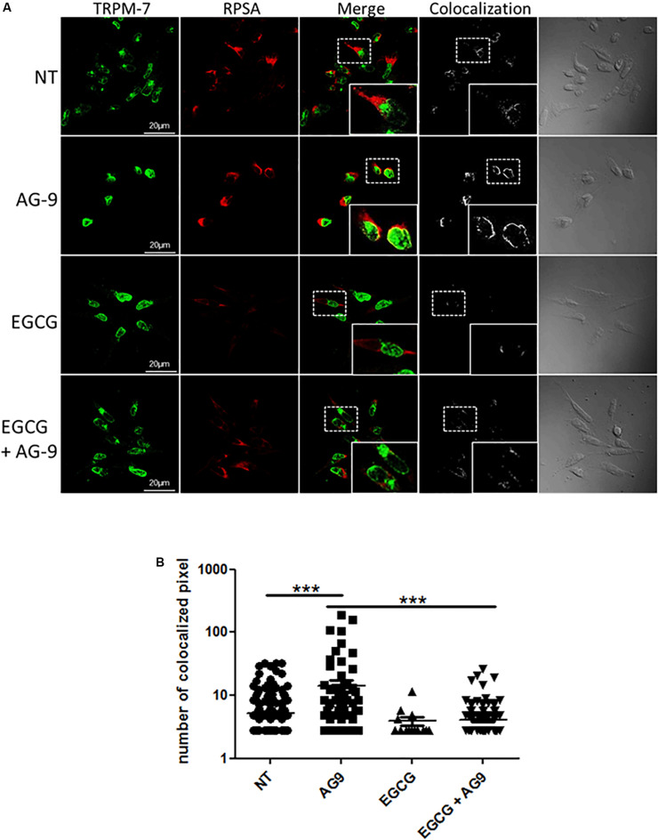
FIGURE 3.
Cellular distribution of TRPM7 and RPSA. (A) MIA PaCa-2 cells were pre-incubated with or without EGCG (10 μM) for 1 h and then with or without AG-9 (10–7 M) for 24 h at +37°C and analyzed by confocal microscopy for TRPM7 and RPSA protein cellular distribution. Colocalization was studied with the Colocalization plugin of ImageJ. Inserts: 2.25× magnification. Scale bar: 20 μm. (B) Quantification of TRPM7/RPSA colocalization pixels in confocal optical sections of MIA PaCa-2 cells in the presence or not of AG-9 (10–7 M) and EGCG (10 μM). Data from one experiment, representative of three independent experiments, are shown. ***P < 0.001 by Mann-Whitney rank sum tests.

