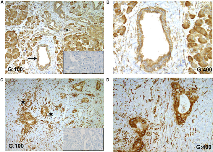
FIGURE 4.
RPSA expression in human PDAC. (A) RPSA is ubiquitously expressed in the normal pancreatic tissue (pancreatic duct and acinar cells, inflammatory, and stromal cells) but no unspecific staining was seen in collagen fibers, black arrows focus on healthy pancreatic ducts. (B) At high magnification, a weak and cytosolic staining was observed in normal duct cells. (C) In PDAC tissue, a high staining was observed in all tumoral cells, and black stars show tumoral glandular structures. (D) At high magnification, a high and cytosolic staining was always observed in tumoral cells. Inserts: RPSA staining was not apparent in the absence of the primary antibody.

