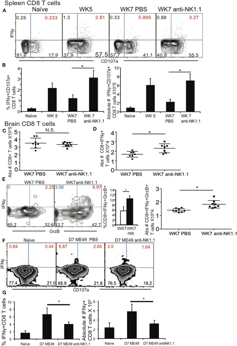
Figure 3.
NK cells reduce polyfunctional CD8+ T cells by increasing their apoptosis during chronic T. gondii infection. C57BL/6 mice were infected as described and 5 weeks after infection treated with 200 μg anti-NK1.1 or 200 μl 1 X PBS i.p. Brain and spleen cells were harvested and restimulated ex vivo with TLA. (A) Contour plots present spleen CD8+ T cells analyzed for IFNγ+ × CD107a+. (B) Graphs present frequency and absolute # of polyfunctional IFNγ+CD107a+CD8+T cells. Data presented is from 1 experiment repeated independently 2 times with n = 5 mice per group. (C) Graphs present pooled data from 2 independent experiments of absolute numbers of brain CD8+ T cells with or without anti-NK1.1 treatment starting at wk 5 post infection. (D) Graphs present pooled data from 2 independent experiments of absolute numbers of brain IFNγ+ CD8+ T cells from animals treated with 200 μg anti-NK1.1 or 1 X PBS i.p. (E) Contour plots of brain IFNγ+GrzB+CD8+ T cells (red numbers are frequency) are presented. Graphs present the frequency and absolute number of brain polyfunctional (IFNγ+Grzb+) CD8+ T cells from 2 independent experiments with n = 4 mice per group. (F,G) B6 mice were treated or not with 200 μg of anti-NK1.1 starting at D-1 then infected with 10 cysts of ME49 strain i.g. Mice were treated every other day with anit-NK1.1. On day 7 post infection animals were sacrificed and spleen cells isolated then stimulated with TLA and stained for polyfunctionality (IFNγ+CD107a+). (F) Contour plots present CD8+ T cells stained for IFNγ+ × CD107a+ during acute T. gondii infection. (G) Graphs present pooled data from 2 experiments showing frequency (%) and absolute number of IFNγ+ CD8+ T cells in spleen. N = 4 mice per group. All graphs are mean ± SD. *denotes significance with a p ≤ 0.05.

