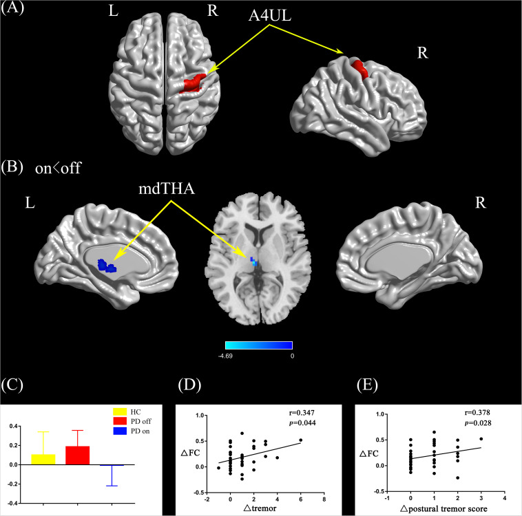
FIGURE 3.
(A) The location of the right upper limb region (red color) based on the Brainnetome Atlas template. (B) Brain region (left mdTHA) showed a significant difference with the right upper limb region in functional connectivity between PD on and PD off (paired t-test, voxel-level p < 0.001, cluster-level p < 0.05, GRF correction), and the cold color indicates decreased functional connectivity in PD on state compared with PD off state (PD on < PD off). (C) FC value histogram for the left mdTHA in the three groups (HC, PD off, PD on). (D,E) ΔFC between the right upper limb region and the left mdTHA show a significant positive correlation with the Δtremor scores of the left upper limb (D) and Δpostural tremor scores of the left upper limb (E). A4UL, area 4 upper limb region; mdTHA, medial dorsal thalamus; ΔFC, difference in functional connectivity between PD on and off states; Δtremor, improvement in symptom scores on UPDRS-III items 20c and 21b (left upper limb); Δpostural tremor score, improvement in symptom scores on UPDRS-III item 21b (left upper limb); L/R, left/right.

