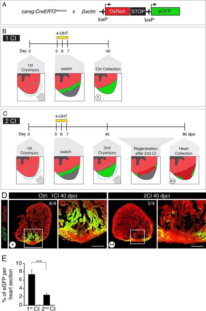
Figure 2.
Repeated cryoinjuries target the same part of the zebrafish heart. (A) Schematic representation of the transgenic fish lines used for the cell-lineage tracing experiment. (B, C) Experimental designs. (B) The strategy to label the regenerated myocardium after cryoinjury (CI). The entire myocardium expresses ßactin:DsRed. careg:Cre-ERT2 is activated in the peri-injury zone in regenerating cardiomyocytes. Treatment with 4-hydroxytamoxifen (4-OHT) for 2 days starting at 5 dpci (days post-cryoinjury) results in Cre-loxP recombination that leads to eGFP expression in the new myocardium, as assessed at 40 dpci. (C) The strategy to assess if the regenerated myocardium is damaged by the subsequent cryoinjury. At 40 dpci, another cryoinjury is performed, and hearts are analyzed after subsequent 40 days. (D) Cross-sections of zebrafish hearts at 40 dpci after one (*) or two (**) cryoinjuries (CIs). The first regenerated myocardium is labelled by eGFP. The second cryoinjury destroyed this regenerated tissue, as revealed by the loss of the majority of the eGFP-positive myocardium. The number in the upper right corner of each image represents the fraction of analyzed fish with the displayed phenotype. Scale bar = 100 µm. (E) Histogram showing the proportion of eGFP-labelled myocardium relative to the ventricular area after one or two cryoinjuries. The 2nd cryoinjury leads to a significant decrease of 4-OHT-induced eGFP expressing cells compared to hearts after one cryoinjury. N = 4. In this and all subsequent figures, frames depict the areas that are shown at higher magnification to the right of each image.

