Figure 3.
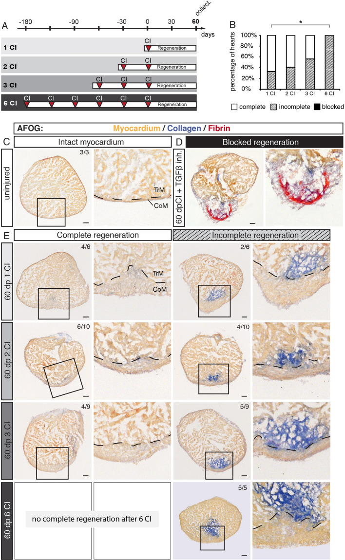
The capacity of complete regeneration is limited after six cryoinjuries. (A) Experimental design showing a schedule of cryoinjuries that were performed every month in the same animals. After the last cryoinjury, hearts were allowed to regenerate for 60 days. (B) Histograms representing the percentage of zebrafish hearts with complete (white), incomplete (gray) or blocked (black) regeneration after one, two, three or six cryoinjuries (CIs). N ≥ 5. *P < 0.05, Student t-test. (C–E) Histological staining of cross-sections with AFOG reagent showing the myocardium (beige), fibrin (red) and collagen (blue). (C) Uninjured hearts do not contain collagen (blue) in the myocardium. (D) An example of blocked regeneration at 60 dpci, following inhibition of the TGF-ß signaling pathway with the chemical antagonist SB431542. Blocked regeneration is featured by the presence of fibrin around the wound, and a lack of myocardium in the wounded area. (E) Representative sections of hearts exhibiting complete and incomplete regeneration at 60 dpci after one, two, three or six cryoinjuries. Dashed line in images at higher magnification marks the junctional region between the trabecular (TrM) and compact (CoM) myocardium. The number in the upper right corner of each image represents the fraction of analyzed fish with the displayed phenotype. Scale bar = 50 µm.

