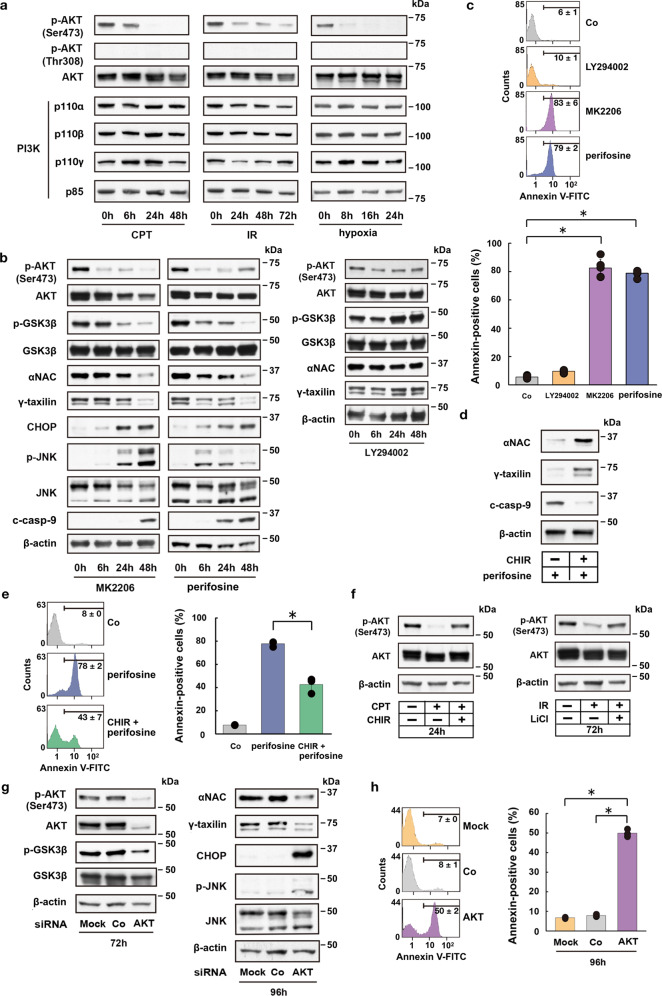
Fig. 2. AKT modulates GSK3β-dependent ER-stress response pathway.
a Western blot analysis shows kinetic changes of Ser473 phosphorylated AKT (p-AKT) and PI3K component (p110α, p110β, p110γ, p85) protein levels in CPT- or IR-treated, or hypoxic HeLa S3 cells. b Western blot analysis shows kinetic changes of p-AKT (Ser473), p-GSK3β (Ser9), αNAC, γTX, and ER-stress response-related proteins in HeLa S3 cells after addition of AKT (MK2206 or perifosine) or PI3K (LY294002) inhibitors. c FACS analysis shows accelerated apoptotic cell death by AKT inhibition (MK2206 or perifosine), but not by PI3K inhibition (LY294002) in HeLa S3 cells. Bars, mean ± s.e.m.; n = 6 independent experiments; *P < 0.001, Tukey–Kramer test. d Western blot analysis for αNAC, γTX and c-casp-9 following GSK3β inhibition (CHIR) in perifosine-treated HeLa S3 cells. e FACS analysis shows suppression of apoptotic cell death following GSK3β inhibition (CHIR) in perifosine-treated HeLa S3 cells. Bars, mean ± s.e.m.; n = 3 independent experiments; *P < 0.001, Tukey–Kramer test. f Western blot analysis shows feedback regulation of p-AKT after GSK3β inhibition (CHIR or LiCl) in CPT- or IR-treated HeLa S3 cells. g Western blot analysis shows AKT siRNA-induced suppression of p-GSK3β (72 h), αNAC (96 h), and γTX (96 h) expression, and induction of CHOP and p-JNK expression (96 h) in HeLa S3 cells. h FACS analysis shows AKT siRNA-induced apoptotic cell death in HeLa S3 cells. Bars, mean ± s.e.m.; n = 3 independent experiments; *P < 0.001, Tukey–Kramer test.

