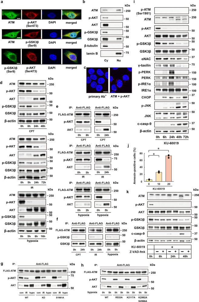
Fig. 3. Cytoplasmic ATM serves as a platform on which AKT protein is inactivated under ER stress.
a Fluorescence confocal microscopy shows subcellular localization of ATM, p-AKT and p-GSK3β in HeLa S3 cells. Scale bar = 5 μm. b Western blot analysis for AKT, p-AKT, GSK3β and p-GSK3β proteins in cytoplasmic (Cy) or nuclear (Nu) cell lysates from HeLa S3 cells. c Proximity ligation assay (PLA) reveals multiple PLA puncta in the cytoplasm and to lesser extent in the nuclear periphery of HeLa S3 cells. PLA without primary antibodies (primary Ab−) or with ATM- and p-AKT-specific antibodies (ATM + p-AKT). Scale bar = 10 μm. d Western blot analysis for 293T cells shows time-dependent downregulation of p-AKT and p-GSK3β proteins after treatment with CPT (1 μM) or IR (20 Gy), or under hypoxic stress. e Immunoprecipitation assay shows depletion of p-AKT in FLAG-tagged ATM precipitates from 293T cells treated with CPT (24 h) or IR (24 h), or from cells under hypoxia (5 h). f Immunoprecipitation assay for GSK3β and p-GSK3β proteins in FLAG-tagged ATM precipitates from 293T cells after treatment with CPT or IR, or from cells under hypoxia. g Immunoprecipitation assay for AKT and p-AKT proteins in FLAG-tagged wild-type (WT) or mutant (KD or S1981A) ATM precipitates before (con) and after ER stress (IR or hypo). h Immunoprecipitation assay for AKT and p-AKT proteins in FLAG-tagged wild-type (WT) or mutant (R533A, K2117A, or K2992A/S2996A; Supplementary Table 1) ATM precipitates from control (0 h) and hypoxic (5 h) 293 T cells. i Western blot analysis shows ER-stress response-mimicking changes in protein expression levels caused by the ATM-specific inhibitor KU-60019 in HeLa S3 cells. j Bar graph shows dose-dependent (0, 10, or 20 μM) enhancement by KU-60019 of apoptotic cell death. Bars, mean ± s.e.m.; n = 5 independent experiments; *P < 0.001, Tukey–Kramer test. k Western blot analysis shows caspase-dependent (Z-VAD-fmk-sensitive) degradation of ATM in KU-60019 (20 μM)-treated HeLa S3 cells, opposing to caspase-independent (Z-VAD-fmk-insensitive) downregulation of p-AKT and p-GSK3β.

