
Figure 8.
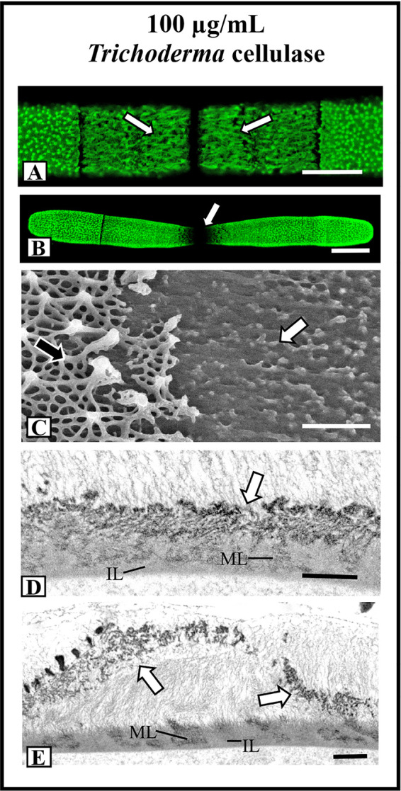
Effects of cellulase treatments on lattice architecture. (A) After 12 h of incubation in 100 µg/ml Trichoderma cellulose, the lattice transforms into elongate fibers (arrows). OGA7-13488 label, confocal laser scanning microscopy (CLSM) image. Bar, 8 µm. (B) After 36 h of treatment in cellulose, the cell shape transforms whereby the isthmus narrow and no lattice is apparent (arrow). OGA7-13488 label, CLSM image Bar, 17 µm. (C) Field emission scanning electron microscopy (FESEM) image of the cell wall surface of a cell treated with cellulose for 36 h. Note the removal of most of the lattice (white arrow). The pre-existing lattice is also observable (black arrow). Bar, 1 µm. (D) Transmission electron miscroscopy (TEM) image of the wall of a cell treated with cellulase for 24 h. Note that the lattice is not solid (arrow) but consists of punctate components. The medial (ML) and inner (IL) layer are observable. Bar. 750 nm. (E) TEM image of the lattice remnant peeling off the cell wall (arrows) in a cell treated with cellulose for 36 h. The medial (ML) and inner (IL) layer are observable. Bar, 900 nm.