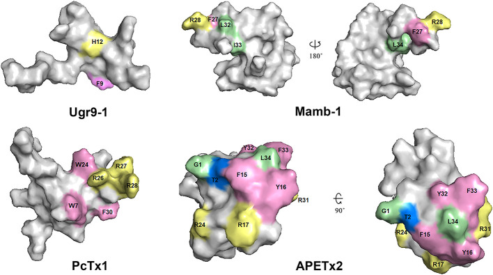
Figure 11.
Spatial structure of toxins Ugr9-1 (PDB 2LZO), Mamb-1 (PDB 5DZ5), PcTx1 (PDB 2KNI), and APETx2 (PDB 2MUB). Marked residues play an important role in the activity of toxins on ASICs in accordance with scanning mutagenesis experiments. Basic, aromatic, and hydrophobic residues are indicated by light yellow, light lilac, and light green colors, respectively.

