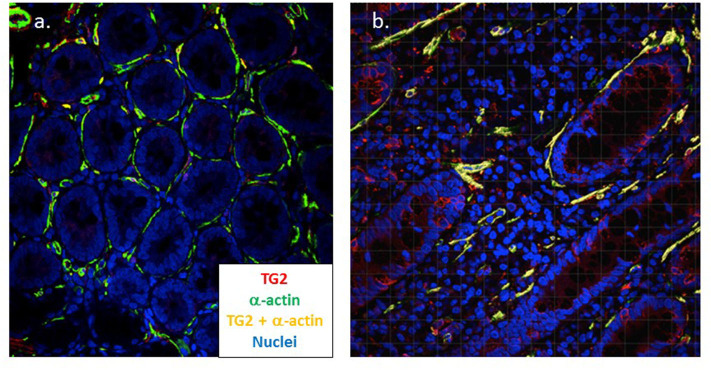
Figure 3.
Myofibroblasts strongly co-express TG2 and α-actin in coeliac disease. Dual color confocal microscopy demonstrates that intestinal myofibroblasts stain positive for α-actin (green) in healthy control tissue (n = 5) (a). In active coeliac disease (n = 11) (b) these cells upregulate TG2 (red) and significant co-expression is apparent (yellow) (Cooper et al., manuscript in preparation). Original magnification x40.

