
Figure EV4. Related to Figs 5 and 6. Reappearance of MRC subunits and COX7A2L and assembly kinetics into respirasomes upon reversible inhibition of mitochondrial translation.
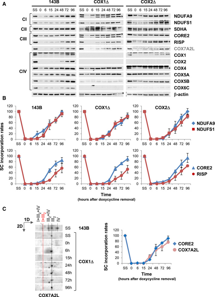
Control 143B cells, and COX1Δ and COX2Δ cybrids were cultured for 6 days in the presence of 15 μg/ml doxycycline and harvested at different time points (in hours) after doxycycline removal. Untreated cells (SS) were included as a positive control.
-
AWhole‐cell extracts were subsequently analyzed by SDS–PAGE and immunoblotting with the indicated antibodies. β‐actin was used as a loading control. Asterisks indicate the matching bands recognized by the antibodies.
-
BMitochondria were extracted with a digitonin‐to‐protein ratio of 2:1 g/g and analyzed by BN–PAGE. The assembly dynamics of CI subunits NDUFA9 and NDUFS1, involved in early and late steps of CI assembly, and of CIII subunits CORE2 and RISP, involved in early and late steps of CIII assembly, were compared in the three cell lines.
-
CAssembly kinetics of COX7A2L relative to CORE2 in the COX1Δ cybrids. The directions of electrophoresis on 2D‐BN/SDS–PAGE gels are indicated with arrows. I+III2+IV, SCs containing CI, CIII, and CIV; I+III2, SC containing CI and CIII; III2+IV, SC containing CIII and CIV; III2, CIII dimer; IV, CIV monomer.