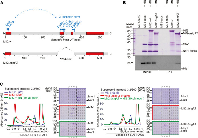
Figure 6. Mif2 auto‐inhibition depends on its Cse4 and DNA‐binding motifs.

- Schematic introduction of Mif2wt and Mif2‐ΔsigAT in which its Cse4 and DNA‐binding motifs have been removed. Cross‐linked lysin residues that connect the N‐terminus with residues within the signature motif or AT‐hook of Mif2 in isolation are depicted in blue and were the basis to create the Mif2‐ΔsigAT mutant.
- Mif2‐Flag‐wt or Mif2‐Flag‐ΔsigAT directly from insect cell lysate was immobilized on M2 Flag Agarose, washed, and incubated with Mtw1‐Nnf1 complex (MN). After washing, bound complexes were eluted by adding 3xFlag peptide. Western blotting against the His‐tag on Nnf1 confirmed increased binding of MN to Mif2‐ΔsigAT, already visible in the Coomassie‐stained SDS–PAGE gel.
- Analytical SEC chromatograms and corresponding Coomassie‐stained SDS–PAGE gels of Mif2‐wt or Mif2‐ΔsigAT and MN alone and in combination at 10 μM concentration. All combinations were incubated for 1 h at 4°C prior to the run. MN (blue), Mif2 (red), combination (green). Note that the same MN elution profile and SDS–PAGE (upper panel) are displayed in both sets to improve clarity. Dashed boxes highlighting corresponding fractions were included to improve comparability.
