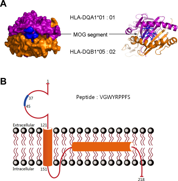
Figure 3.

Computational docking of the MOG segment VGWYRPPFS to the HLA-DQB1*05:02 heterodimer and structure of the MOG protein. (A) In silico docking simulation resulted in the placement of the MOG segment within the peptide-binding groove between HLA-DQB1*05:02 and HLA-DQA1 with docking scores (∆G) of −10.4 kcal/mol. Mesh on the surface of HLA indicates close contact between the atoms and MOG protein segment. The ribbon structure shows the ligand-binding domains. (B) The structure of the MOG protein and the blue sections indicate the location of the peptide segment. HLA, human leucocyte antigen; MOG, myelin oligodendrocyte glycoprotein.
