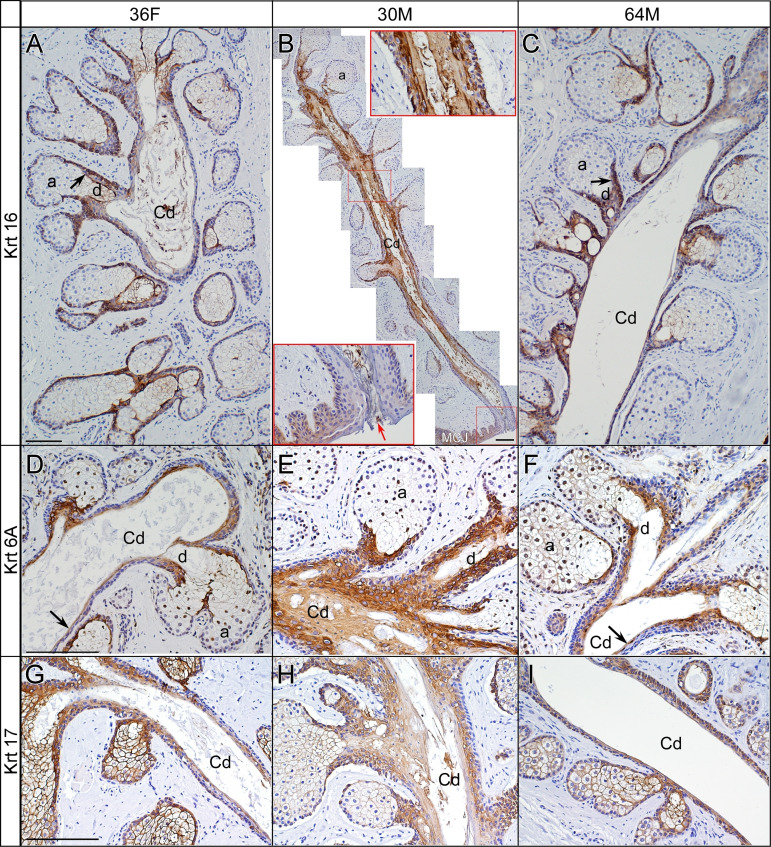
Figure 3.
Expression of Krt16, Krt6a and Krt17 in MGs of an older donor (64M) and of the two young donors, one with normal-appearing MGs (36F) and the other with evidence of ductal obstruction (30M). (A–C) Krt16 protein was mostly distributed in MG ductules and decreased in the MG central duct of 36F (A) and 64M (C) as compared with Krt16 expression in 30M (B). The central ductal cells in 30M showed Krt16 overexpression (B, montage and inset on the top right) and an orifice ‘plug’ (B, red arrow in bottom left inset). (D–F) Krt6a expression was similar to Krt16 expression in the MG ductules of 36F (D) and 64M (F). Krt6a overproduction was seen in the central duct of 30M (E). (G–I) Krt17 was ubiquitously expressed in the MGs of 30M, 36F and 64M, with a higher level in the epithelial cells of ductules. Krt17 overexpression was noted in the central duct of 30M, but not in the central duct of 36F and 64M (H). Scale bar denotes 100 µm. 36F, 36-year-old female; 30M, 30-year-old male; 64M, 64-year old male; a, acinus; Cd, central duct; d, ductule; MG, meibomian gland.

