Figure 4.
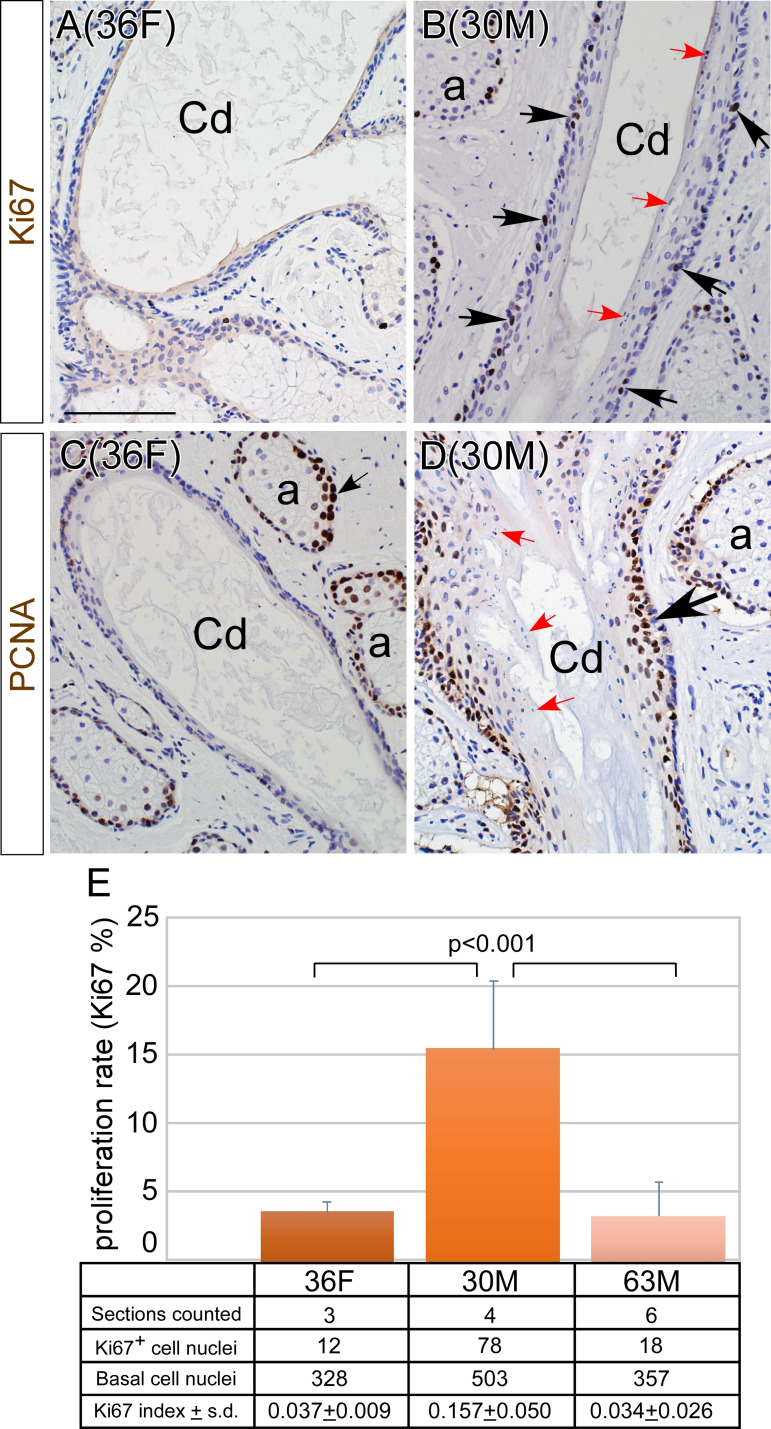
Expression of cell proliferation markers in MGs of the donor with ductal obstruction (30M), the donor with normal-appearing MGs (36F) and one of the older donors (63M). (A, B) Ki67 expression. Ki67-positive cells were rarely identified in the basal epithelia of the central ducts in 36F (A). In contrast, many Ki67-positive cells were noted in the basal epithelia of 30M (B, black arrows). (C, D) PCNA expression was found in the nuclei of the acinar basal epithelium of 36F (C, arrow), and expression was diminished in cells differentiating into meibocytes. PCNA-expressing cells were rarely detected in the MG central duct in 36F (C), whereas in 30M PCNA-positive cells were abundant in the epithelia of the central duct (D, black arrow). In 30M, cell nuclear fragments were retained in the superficial layer of the MG central duct (B and D, red arrows). (E) Ki67 labelling index in MG ductal basal epithelia. Cell proliferation rate in ductal basal epithelia was higher in 30M than in 36F and 63M (p<0.001). Scale bar denotes 100 µm. 36F, 36-year-old female; 30M, 30-year-old male; 63M, 63-year old male; a, acinus; CD, central duct; D, ductile; MG, meibomian gland; PCNA, proliferating cell nuclear antigen.

