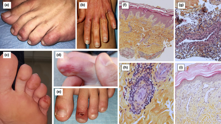
Figure 1.

Skin lesions (a: Patient 7; b: Patient 9; c: Patient 10; d: Patient 4; e: Patient 3); Lesional skin biopsies, Haematoxylin, Eosin and Saffron (HES) (f: lymphohistiocytic infiltrate around vessels and eccrine glands, ×4 magnification; g: Angiocentric lymphohistiocytic infiltrate in superficial dermis, ×40 magnification; h: Angiotropism, ×40 magnification; i: Papillary dermis oedema, capillar ectasia and endothelial swelling, ×20 magnification).
