
Figure 2.
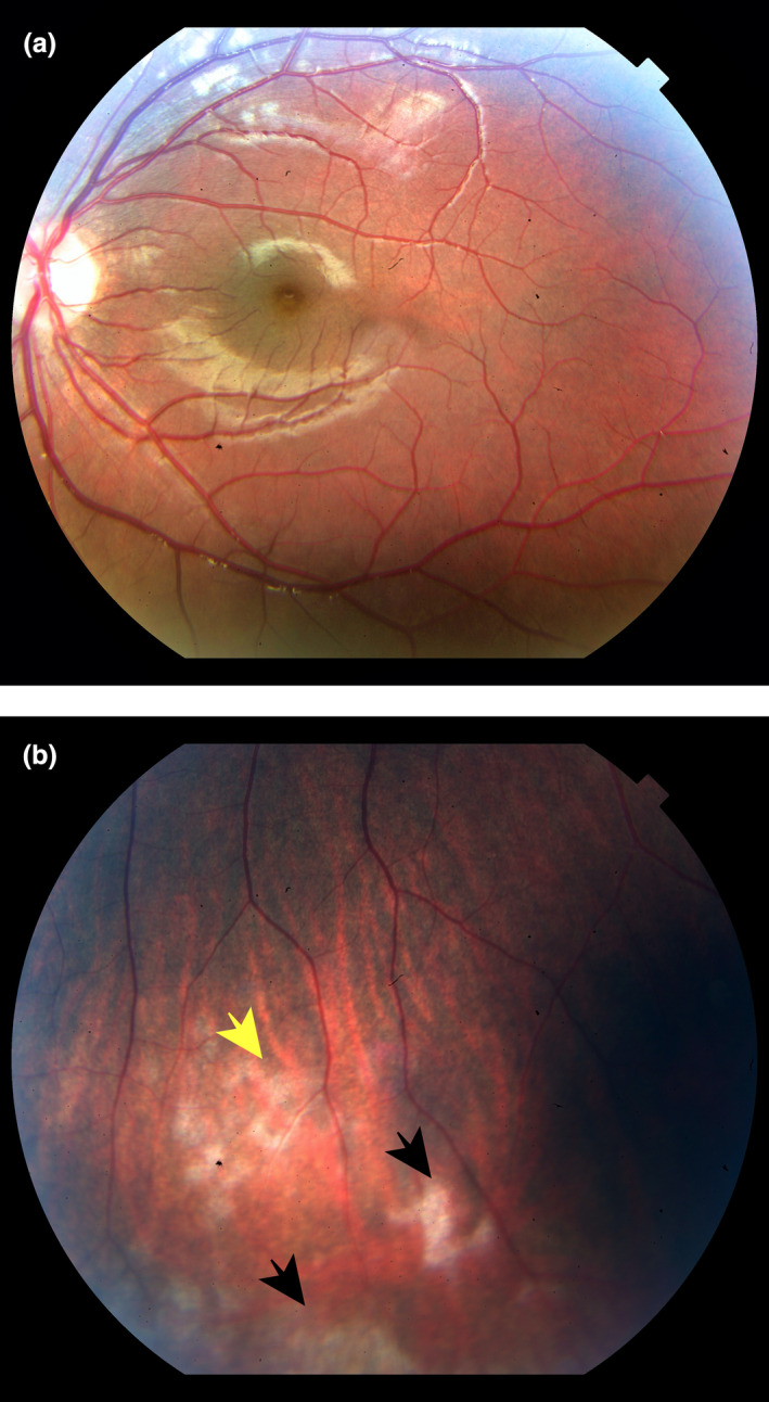
Funduscopic examination. The image does not show significant pathologic features on right eye (a). Retinal vasculitis with perivascular infiltrate (yellow arrow) and retinal exudates (black arrows) on retinal equator of left eye (b).
Official websites use .gov
A
.gov website belongs to an official
government organization in the United States.
Secure .gov websites use HTTPS
A lock (
) or https:// means you've safely
connected to the .gov website. Share sensitive
information only on official, secure websites.
Funduscopic examination. The image does not show significant pathologic features on right eye (a). Retinal vasculitis with perivascular infiltrate (yellow arrow) and retinal exudates (black arrows) on retinal equator of left eye (b).