Figure 1.

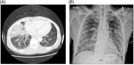
Computed tomography chest imaging showing a right lung middle lobe consolidation with bilateral ground glass opacity (A) in patient 1 and an anterior posterior chest X‐ray (B) showing bilateral lower lobe infiltrates

Computed tomography chest imaging showing a right lung middle lobe consolidation with bilateral ground glass opacity (A) in patient 1 and an anterior posterior chest X‐ray (B) showing bilateral lower lobe infiltrates