Figure 1.
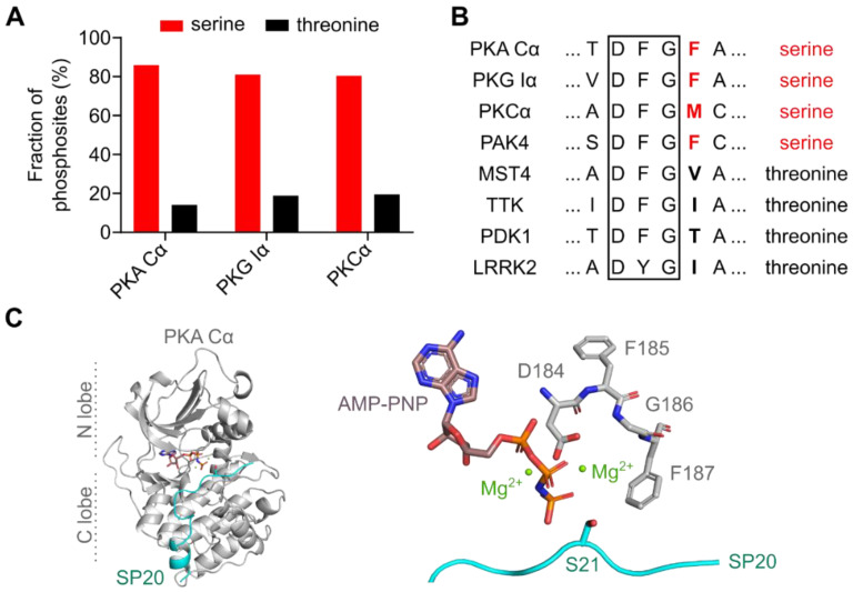
PKA prefers serine over threonine as a phosphoryl acceptor residue. (A) The AGC kinases PKA, PKG, and PKC are serine-specific. Bar diagram showing the fraction of annotated phosphosites for serine and threonine phosphoryl acceptors. Data were obtained from the PhosphoSitePlus® database v6.5.9.1 [23]. (B) Alignment of the DFG motif (black box) and the DFG+1 residues (bold letters) of human Ser/Thr protein kinases that prefer either serine (red) or threonine (black) as phosphoryl acceptor. The alignment was generated with Clustal Omega [24]. UniProt IDs: P17612 (PKA Cα); Q13976 (PKG Iα); P17252 (PKCα); O96013 (PAK4); Q9P289 (MST4); P33981 (TTK); O15530 (PDK1); Q5S007 (LRRK2). (C) Crystal structure of the murine PKA Cα subunit with AMP-PNP, Mg2+, and SP20 bound (PDB code: 4DG0) [25]. A zoomed view (right panel) shows the DFG motif (residues 184-186) and the DFG+1 residue (F187) interacting with Mg2AMP-PNP and the substrate SP20. S21 is the phosphoryl acceptor residue of SP20. All structure images were generated using the PyMOL Molecular Graphics System (Version 2.2.2; Schrödinger, LLC, New York, NY, USA).

