Figure 3.
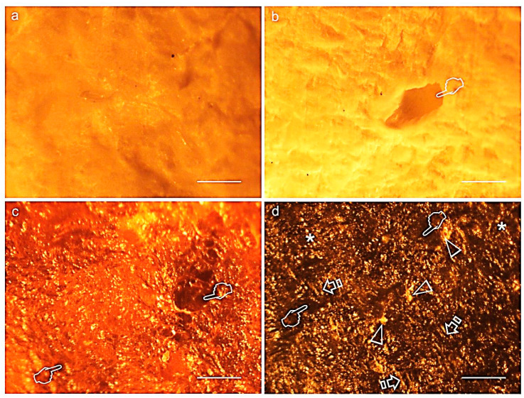
Representative light micrographs of surfaces of Derma Fina membrane at the outer (a), inner (b) surfaces before immersion and at the inner surfaces after trypsin (c) and PBS (d) immersion, at 21 d of storage. Single arrows indicate bundles of collagen. Both membrane faces (b,c) showed the typical crumpled appearance of immersed membranes. Large diameter pores (wider than 100 µm) were observed at the surface of the membrane (pointers). Shorter and thinner fibers were seen after 21 d in trypsin, and large and thicker fibers characterized the membrane surface after biodegradation in PBS (asterisks). A compact collagen arrangement was interrupted by clusters of circular discontinuities, probably mineral depositions (arrow heads). Scale bar is 1000 µm in (a,c,d); and 2000 µm in (b).

