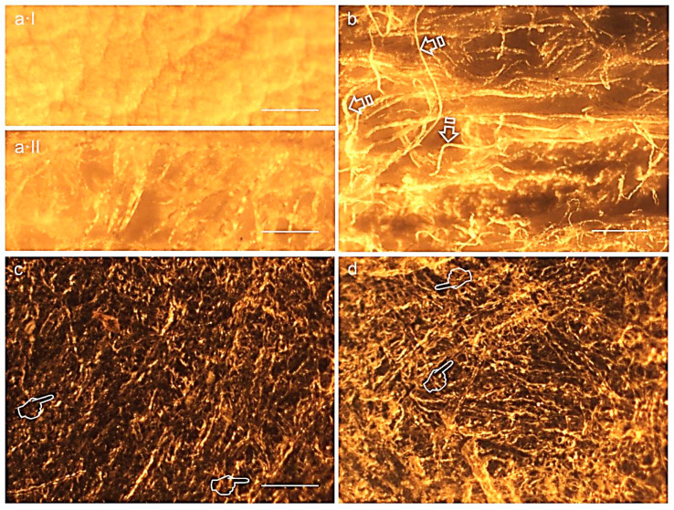
Figure 4.
Representative light micrographs of surfaces of Evolution standard membrane at the outer (a·I), inner (a·II, b) surfaces before immersion and at the inner surfaces after trypsin (c) and PBS (d) immersion, at 21 d of storage. The ondulating collagen fibers and microfibrils were arranged in stacked layers, which were parallel to the membrane surface (single arrows). Collagen fibrils formed fine, loosely arranged, undulating collagen bundles. The packing of fibrils into fibers was not very tight, resulting in single fibrils or small bundles interconnecting larger bundles. Isolated fibrils running crosswise formed a loose meshwork overlying the bundled fibrils. Multiple grooves, pits and pores (pointers) were seen at the inner surface of the membrane. Scale bar is 1000 µm in (a·I), (a·II), (c), (d) and 2000 µm in (b).

