Figure 1.

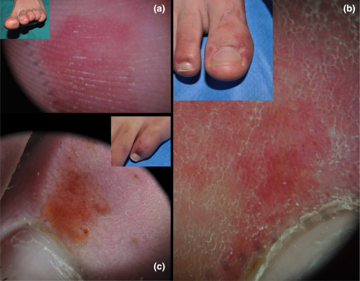
(a) Homogeneous red area on the tip of toe. No globules or reticule are seen. (b) Red background area with purpuric globules on the perionychium. (c) Brown area with reticule and brown globules both within the area and peripherically.

(a) Homogeneous red area on the tip of toe. No globules or reticule are seen. (b) Red background area with purpuric globules on the perionychium. (c) Brown area with reticule and brown globules both within the area and peripherically.