
Figure 6.
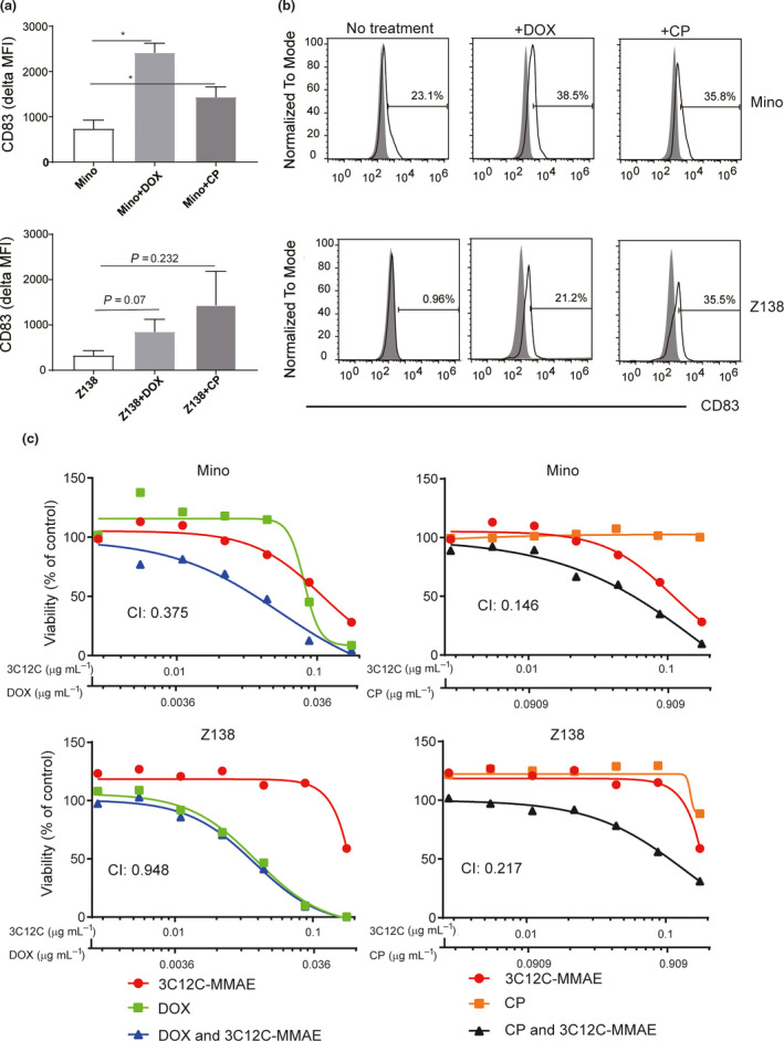
3C12C ADC synergised with doxorubicin/cyclophosphamide. Mino or Z138 cells were cultured in the presence of DOX (0.02 µg mL−1) or CP (0.2 mg mL−1) for 48 h, and cell surface CD83 expression was analysed by flow cytometry. Mean fluorescent intensity ± SEM of three experiments (a) and data from one of three experiments (b) are shown. (c) Mino or Z138 cells were cultured with serially diluted 3C12C‐MMAE (0.176, 0.088, 0.044, 0.022, 0.011, 0.0055, 0.00275 µg mL−1), DOX (0.064, 0.032, 0.016, 0.008, 0.004, 0.002, 0.001 µg mL−1), CP (1.6, 0.8, 0.4, 0.2, 0.1, 0.05, 0.025 µg mL−1) or the combination of 3C12C‐MMAE/DOX (0.176/0.064, 0.088/0.032, 0.044/0.016, 0.022/0.008, 0.011/0.004, 0.0055/0.002, 0.00275/0.001 µg mL−1), and the combination of 3C12C‐MMAE/CP (0.176/1.6, 0.088/0.8, 0.044/0.4, 0.022/0.2 0.011/0.1, 0.0055/0.05, 0.00275/0.025 µg mL−1) for 72 h. CellTiter‐Glo Luminescent cell viability assay was used to determine the killing effect. Data are from one of three independent experiments. The Combination Index (CI) was analysed with CompuSyn.