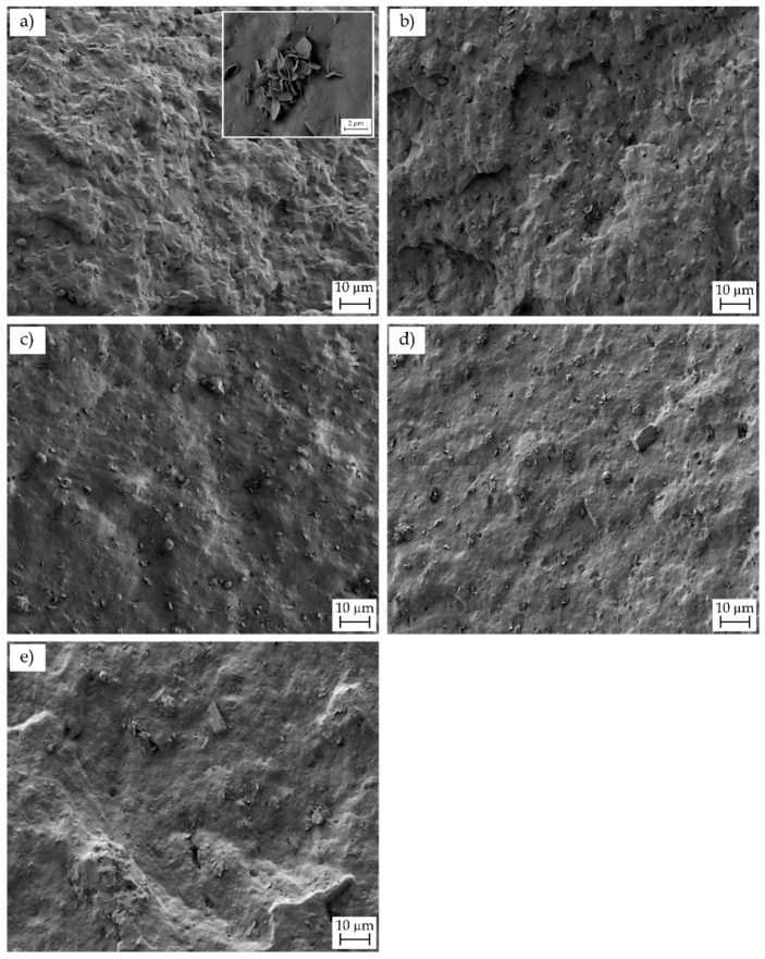
Figure 8.
Field-emission scanning electron microscopy (FESEM) images of the fracture surfaces of the injection-molded poly(3-hydroxybutyrate-co-3-hydroxyhexanoate) [P(3HB-co-3HHx)]/hydroxyapatite nanoparticle (nHA) parts of: (a) neat P(3HB-co-3HHx); (b) P(3HB-co-3HHx) + 2.5 nHA; (c) P(3HB-co-3HHx) + 5 nHA; (d) P(3HB-co-3HHx) + 10 nHA; (e) P(3HB-co-3HHx) + 20 nHA. Images were taken at 500× and with scale markers of 10 µm. Inset image showing the detail of the microparticles was taken at 2500× with scale marker of 2 µm.

