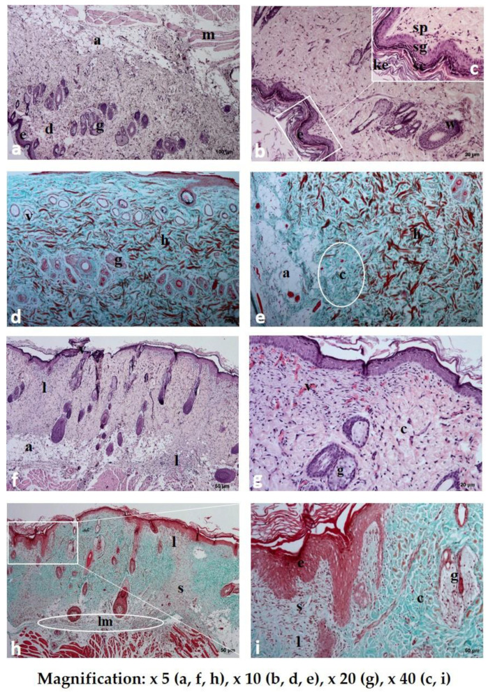
Figure 9.
Histological examination of rat skin after the application of Spongostan (a–e) and placebo film (PVA/HPMC_B/C) (f–i) stained with hematoxylin and eosin (HE a–c,f,g) and Masson’s trichrome (d,e,h,i) staining: e-epidermis; sc-stratum corneum; sg-stratum granulosum; sp-stratum spinosum; ke-keratin; d-dermis; h-hemorrhagic zones; a-adipose tissue; m-muscular cells; g-sebaceous gland; c-collagen fibers; lm-leukocytes and macrophages; s, ”scar.” In the subcutaneous part of rat skin after the application of placebo film (h), a visible layer of inflammatory cells (lm) and a zone with disturbed layers, structurally different from the surrounding, healthy tissues, were observed.

