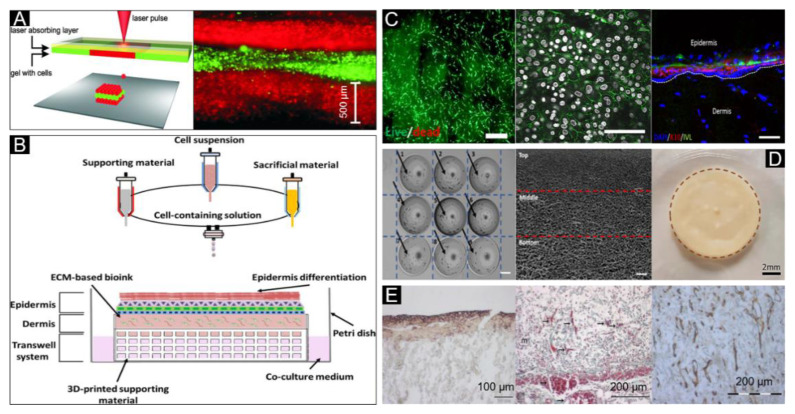
Figure 8.
Shear-thinning bioink for skin bioprinting. (A) Sketch of the laser printing setup (left) and the alternating color-layers of red (keratinocytes) and green (fibroblasts) (right). Reproduced with permission from [83]; copyright Wiley Periodicals, 2012. (B) Extrusion-inkjet printing pattern for fabrication of 3D printed human skin model and its maturation with functional transwell system, all in a single-step process. (C) Image and dead images of the contracted compartments at day 7 to reveal their morphology and viability (left, scale bar: 200 μm), immunofluorescence labelling examination of E-cadherin (middle, white: Nuclei, green: E-cadherin, scale bar: 200 μm), and immunofluorescence analysis of the human skin model (Right, DAPI: Nucleus, K10: Early differentiation marker, and IVL: Involucrin, scale bar: 100 μm). Reproduced with permission from [160]; copyright IOP, 2017. (D) A two-step 3D bioprinting strategy to manipulate the cell distribution (cells indicated by black arrows—homogeneous cell distribution) (Left), pore size distribution with the 3D collagen matrix-hierarchical porous microstructure (middle), and 3D bioprinted pigmented human skin constructs with uniform skin pigmentation (right), pigmented area is enclosed by the brown dotted line. Reproduced with permission from [116]; copyright IOP, 2018. (E) E-cadherin expression (left), blood vessels (arrows) (middle) and collagen IV (right) can be detected in the skin constructs on day 11. Reproduced from [161], under open access license.

