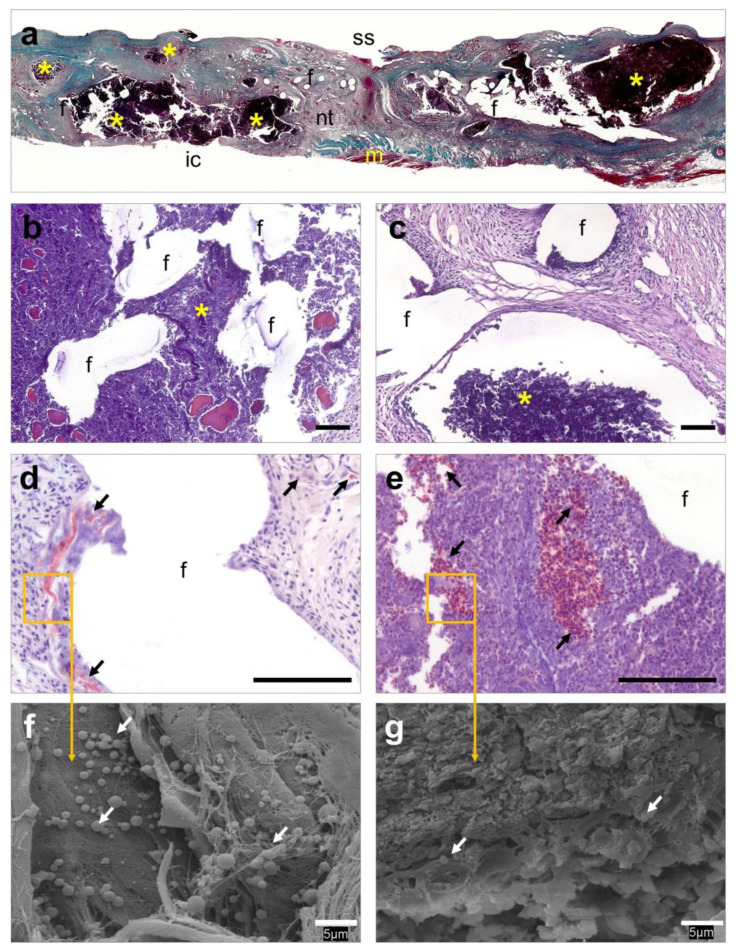
Figure 5.
Histological evaluation of the HApN implants. (a) Panoramic view of an implant displaying several abscesses that disrupt tissue integration (Masson’s trichrome, ×50). (b,c) Detail of different areas of the implant showing a dense connective tissue surrounding the mesh filaments, containing different-sized abscesses next to the mesh filaments (hematoxylin eosin, ×100). (d,e) Presence of bacteria was immunohistochemically confirmed in neoformed tissue and within abscesses (Sa immunostaining, ×320). (f,g) At higher magnification, bacteria were visualized forming colonies firmly adhering to the implant surface and within the abscesses (scanning electron microscopy, SEM, ×2000). Symbols: (ic) intraperitoneal cavity; (f) mesh filaments; (m) muscle; (nt) neoformed tissue; (ss) subcutaneous side; (*) abscess; (→) bacteria. Scale bars represent 100 µm (b–e) and 5 µm (f,g).

