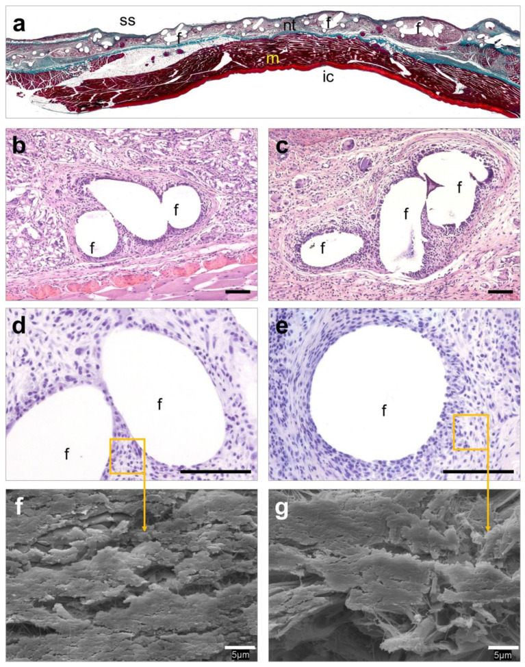
Figure 6.
Histological evaluation of the Rif-HApN implants. (a) Panoramic view of an implant revealing adequate tissue integration (Masson’s trichrome, ×50). (b,c) Detail of different areas of the implant showing a loose neoformed connective tissue surrounding the mesh filaments with no evidence of infection (hematoxylin eosin, ×100). (d,e) No living bacteria were recorded throughout the implant (Sa immunostaining, ×320). (f,g) Scanning electron microscopy (SEM) visualization confirmed the absence of bacteria in these implants (SEM, ×2000). Symbols: (ic) intraperitoneal cavity; (f) mesh filaments; (m) muscle; (nt) neoformed tissue; (ss) subcutaneous side. Scale bars represent 100 µm (b–e) and 5 µm (f,g).

