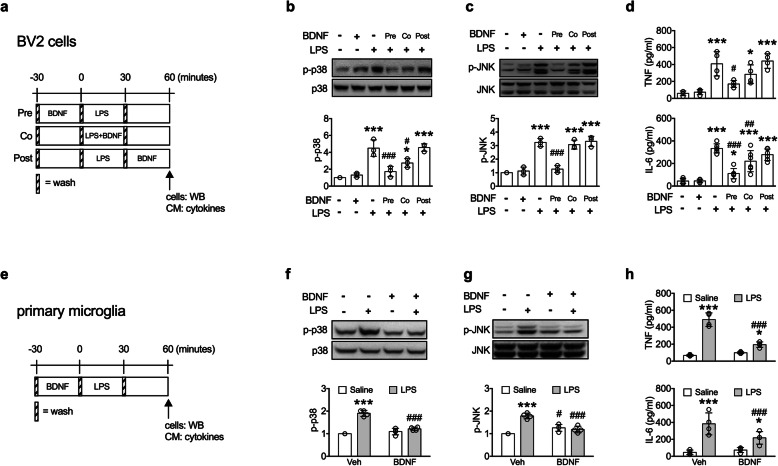
Fig. 4.
LPS-induced microglial activation was blocked by BDNF pre-treatment. a–d BV2 microglial cells. a The experimental timeline. b, c Levels of p-p38 (n = 3) and p-JNK (n = 3) in the BV2 cells. d Levels of TNF (n = 4) and IL-6 (n = 6) in conditioned media. *p < 0.05, ***p < 0.001 versus BDNF(-)LPS(-) group; #p < 0.05, ##p < 0.01, ###p < 0.001 versus BDNF(-)LPS(+) group. e–h Purified primary microglial cells. e The experimental timeline. f, g Levels of p-p38 (n = 4) and p-JNK (n = 4) in the primary microglial cells. h Levels of TNF (n = 4) and IL-6 (n = 4) in conditioned media. *p < 0.05, ***p < 0.001 versus respective Saline group; #p < 0.05, ###p < 0.001 versus respective Veh group (n = 4). Data are presented as mean ± S.D

