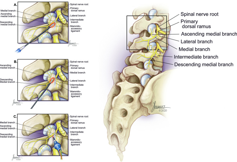
Figure 1.
Representational drawing depicting the lumbosacral facet joints and accompanying neural anatomy. Insets illustrate closeup views of the bony and neural anatomical landmarks and a schematic representation of the effect electrode orientation has on nerve ablation. Artistic renditions by Joe Kanasz (joekanasz@att.net). (A) Parallel insertion of electrodes. Parallel placement may result in a higher likelihood of missing the nerve than with near-parallel orientation. (B) Near-parallel insertion of electrodes. This may result in the highest likelihood of medial branch nerve ablation. (C) Perpendicular insertion of electrodes. This theoretically results in the highest chance of missing the nerve, which may be more likely when the medial branch is entrapped beneath the mammilo-accessory ligament.

