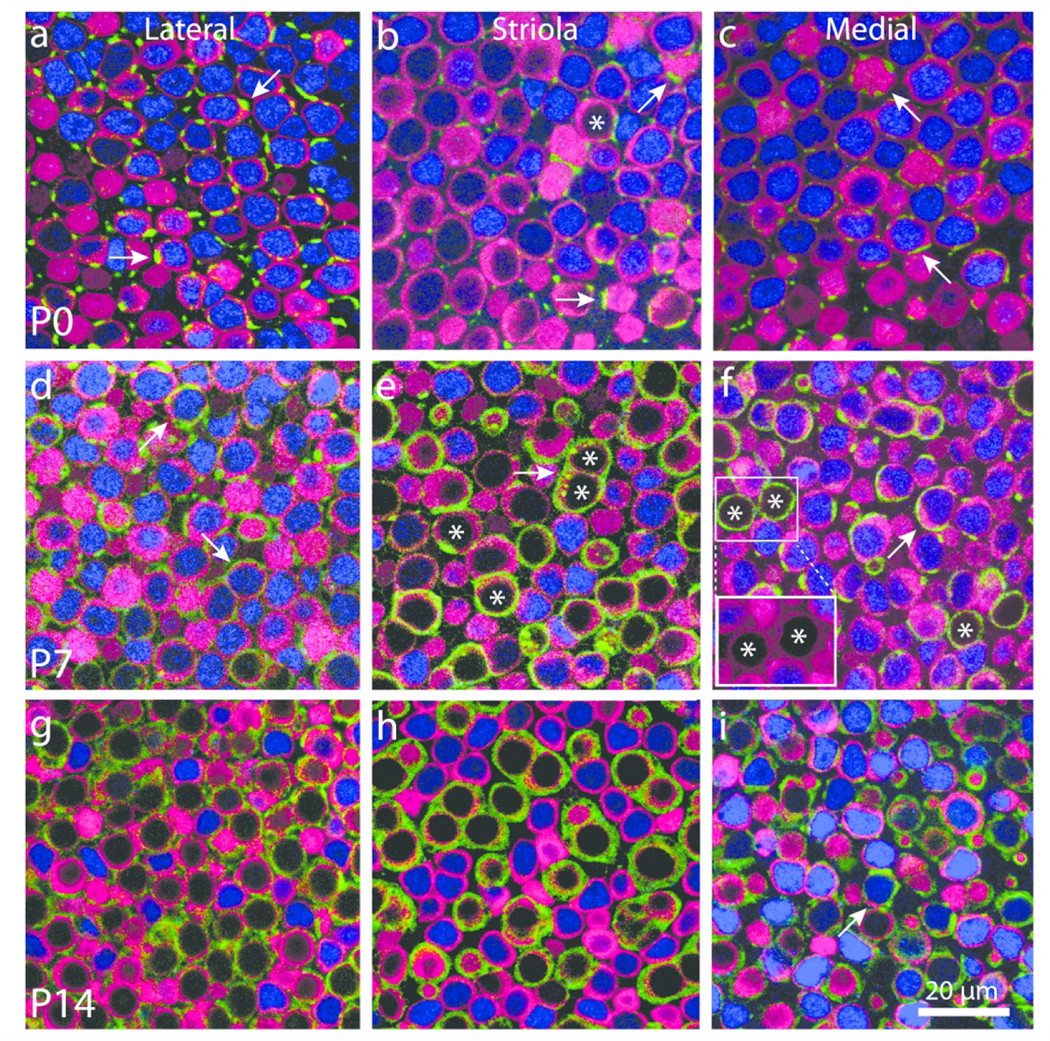
Figure 2.

Calyceal development is not strictly correlated with down-regulation of Sox2 expression. Confocal sections through the sensory epithelia of whole-mount utricles at P0, P7, and P14 immunolabeled for myosin VIIa (hair cells, magenta), β-III tubulin/neurofilament (neurons, green), and Sox2 (blue). Images from the lateral extrastriolar, striolar, and medial extrastriolar region are shown for each age. (a–c) At P0, hair cells that lacked Sox2 expression were present only in the striola (asterisk in panel b). Many hair cells were contacted by thin neuronal processes (green, arrows in a–c), but calyces were very rare. (d–f) Hair cells with Sox2-negative nuclei (examples labeled with asterisk) became more common in the striola and were also observed in the extrastriolar regions. Mature and developing calyces were observed in all regions of the sensory epithelium (arrows in d,f). Such calyces often enclosed hair cells with Sox2-positive nuclei (arrows in d,f). Immunolabeling for hair cells and calyces often overlapped. However, reducing the intensity of the neuronal color channel (green) revealed that all calyces enclosed myosin VIIa-labeled hair cells (red, insert, f). (g–i) Hair cells and calyces were nearly mature in all regions at P14. Most calyx terminals enclosed hair cells that were Sox2-negative, although a few calyces still enclosed Sox2-expressing cells (arrow in i)
