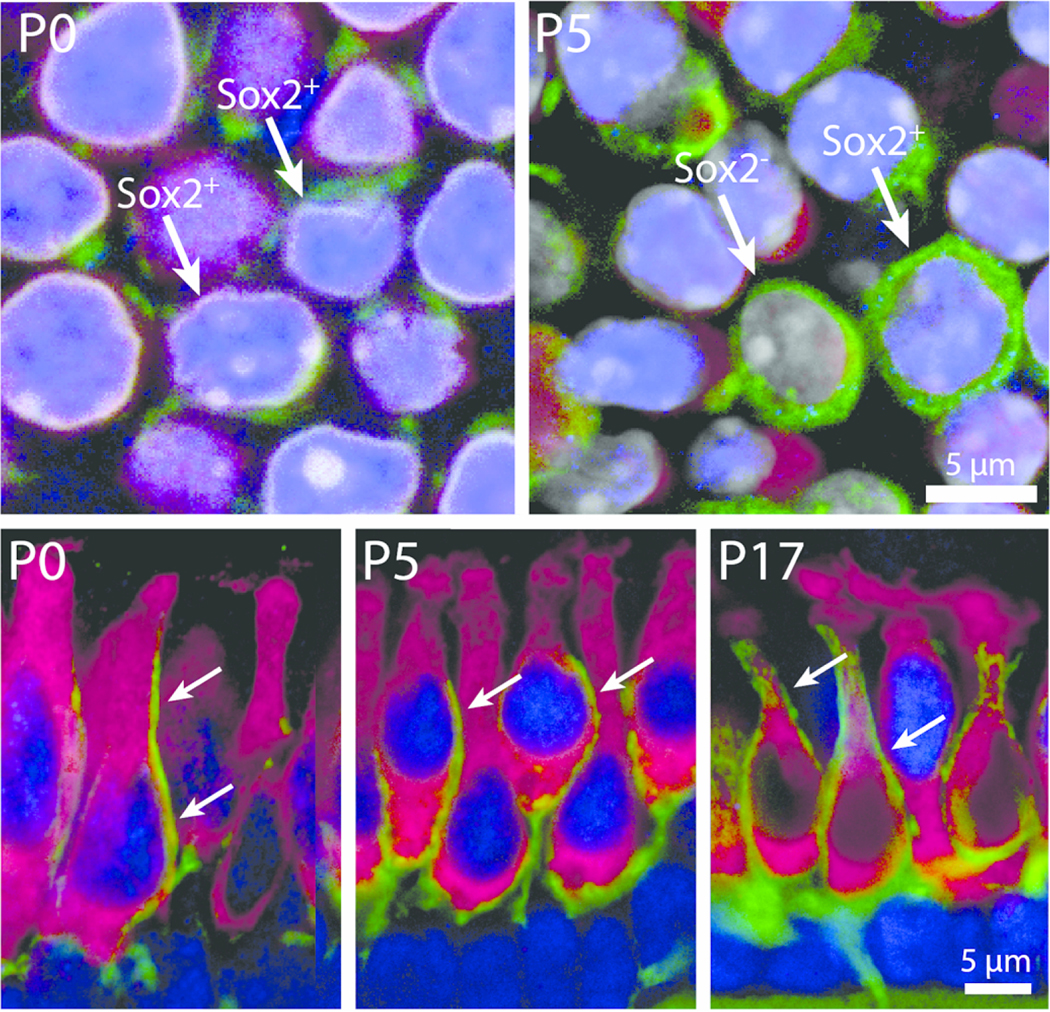
Figure 4.

Postnatal development of calyceal innervation. High magnification images showing the developmental maturation of Type I hair cells and their associated calyx nerve terminals in whole-mount and sectioned utricles. Top row : Z-slice images from the extrastriolar region of the utricle showing hair cells (magenta, myosin VIIa) and developing afferent terminals (green, βIII tubulin/neurofilament). At P0, very few extrastriolar hair cells were enveloped by calyx terminals (green) and nearly all possessed Sox2-positive nuclei (blue, arrows). At P5, however, complete calyces were occasionally observed, and they enclosed either a Sox2-positive hair cell or a Sox2-negative hair cell (arrows). Bottom row : The development of calyx nerve terminals as revealed in cross-sections of frozen specimens. At early postnatal stages (P0 and P5), numerous hair cells in all regions of the sensory epithelium were contacted by long neuronal processes that extended along the apical-basal axis of the hair cell but did not fully enclose the cell body (arrows, P0 and P5). Such processes frequently contacted Sox2-expressing hair cells (blue nuclei). By P17, mature calyx terminals were common in all regions and primarily enclosed Sox2-negative hair cells (P17, arrows)
