Figure 1.
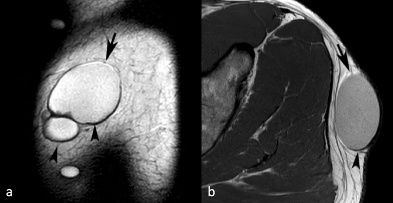
A 44-yr-old male with an epidermoid cyst over the lateral aspect of the left buttock. (a) Sagittal T 2W FSE MR image showing a well-defined multilocular fluid signal intensity lesion with linear hyperintensity around its upper margin (arrow) and hypointensity around its lower margin (arrowheads) due to chemical shift artefact. (b) Similarly, axial PDW FSE MR image showing linear hyperintensity around its anterior margin (arrow) and hypointensity around its posterior margin (arrowhead). PDW FSE, proton densityweighted fast spin echo; T 2W FSE, T 2 weightedfast spin echo.

