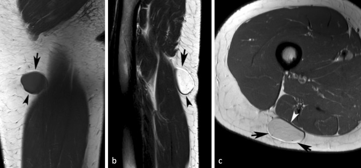
Figure 2.
A 27-yr-old female with a myxoid liposarcoma of the posterior right thigh. (a) Coronal T 1W SE and (b) sagittal T 2W FSE MR images showing a well-defined lesion with linear hyperintensity around its upper margin (arrows) and hypointensity around its lower margin (arrowheads) due to chemical shift artefact. (c) Similarly, axial PDW FSE MR image showing linear hyperintensity around its superficial margin (arrows) and hypointensity around its deep margin (arrowhead). PDW FSE, proton densityweighted fast spin echo; T 1W SE, T 1 weighted spin echo; T 2W FSE,T 2 weightedfast spin echo.

