
Figure 5. Ex vivo [18F]-Alfatide autoradiography and αvβ3 immunofluorescence in liver sections CCl4 and BDL mice.
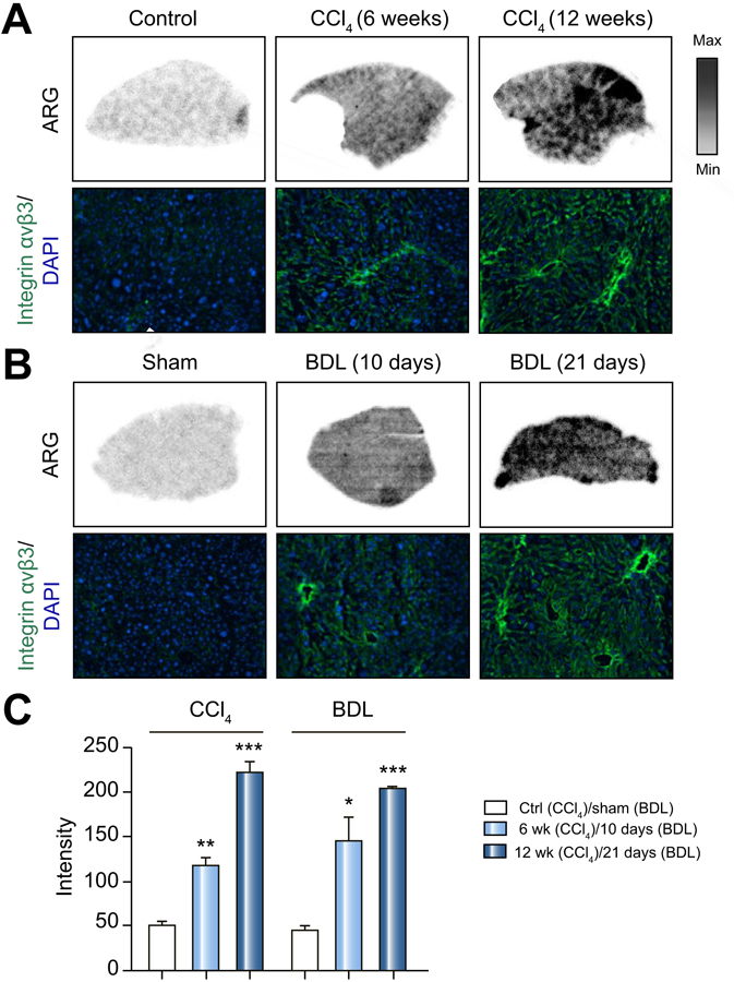
(A) Representative autoradiographic images of liver sections incubated with [18F]-Alfatide for control mice, mild and severe CCl4 mice. Selected area was stained with DAPI (blue) and αvβ3 (Green).
(B) Representative autoradiographic mages of liver sections treated as in (A) for mild and severe BDL mice. Selected area was stained with DAPI (blue) and αvβ3 (Green).
(C) Quantitation of radioactivity as intensity from (A) and (B). Values are mean ±1 SEM. P values are relative to sham.