Figure 7.
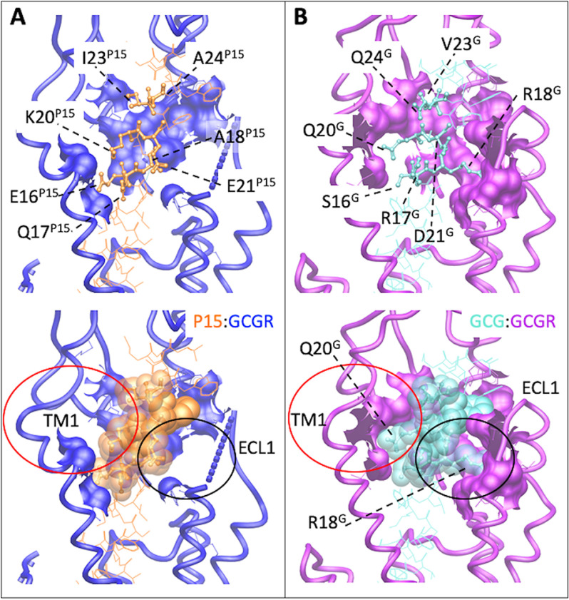
Peptide 15 and glucagon make distinct interactions with GCGR. A, P15–GCGR interface with the divergent peptide amino acids from GCG displayed in x-stick format and conserved residues in wire format. GCGR residues that interact with the nonconserved peptide amino acids are displayed in wire and surface representation. GCGR is blue and P15 orange. The receptor backbone is shown in protein worm representation. B, GCG–GCGR interface with the divergent peptide amino acids from P15 displayed in x-stick format and conserved residues in wire format. GCGR residues that interact with the nonconserved peptide amino acids are displayed in wire and surface representation. GCGR is purple and GCG light blue. The receptor backbone is shown in protein worm representation. Lower panels include cpk representation of the nonconserved peptide residues. The red circle highlights the distinction in interaction surfaces between the top of TM1 and the two peptides. The black circle highlights the distinction in interaction surfaces between ECL1 and the two peptides.

